Atomic structures of two novel immunoglobulin-like domain pairs in the actin cross-linking protein filamin.

Heikkinen OK, Ruskamo S, Konarev PV, Svergun DI, Iivanainen T, Heikkinen SM, Permi P, Koskela H, Kilpeläinen I, Ylänne J, J Biol Chem 284(37):25450-8 (2009) Europe PMC

SASDAS3 – Fila16-17

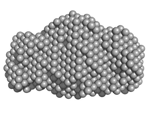
immunoglobulin- like filamin two-domain fragment 16-17
MWexperimental 22 kDa
MWexpected 19 kDa
VPorod 33 nm3
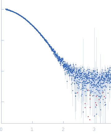
log I(s) 4.15×101 4.15×100 4.15×10⁻¹ 4.15×10⁻²
immunoglobulin- like filamin two-domain fragment 16-17 small angle scattering data  s, nm-1
ln I(s)
immunoglobulin- like filamin two-domain fragment 16-17 Guinier plot ln 4.15×101 Rg: 1.9 nm 0 (1.9 nm)-2 s2
(sRg)2I(s)/I(0)
immunoglobulin- like filamin two-domain fragment 16-17 Kratky plot 1.104 0 3 sRg
p(r)
immunoglobulin- like filamin two-domain fragment 16-17 pair distance distribution function Rg: 1.9 nm 0 Dmax: 6 nm

Data validation


Fits and models


log I(s)
 s, nm-1
immunoglobulin- like filamin two-domain fragment 16-17 GASBOR model

Synchrotron SAXS data from solutions of Fila16-17 in 100 mM NaCl 10 mM dithiothreitol 20 mM Tris, pH 8 were collected on the EMBL X33 beam line at the DORIS III, DESY storage ring (Hamburg, Germany) using a MAR 345 Image Plate detector (I(s) vs s, where s = 4πsinθ/λ, and 2θ is the scattering angle). at 10°C. The data were normalized to the intensity of the transmitted beam and radially averaged; the scattering of the solvent-blank was subtracted.

Wavelength = UNKNOWN. Sample detector distance = UNKNOWN. X-ray Exposure time = UNKNOWN. Number of frames = UNKNOWN. Concentration = UNKNOWN

Tags: X33
immunoglobulin- like filamin two-domain fragment 16-17 (Fila16-17)
Mol. type   Protein
Organism   Escherichia coli
Olig. state   Monomer
Mon. MW   18.7 kDa
Sequence   FASTA