Structural basis of myelin-associated glycoprotein adhesion and signalling.

Pronker MF, Lemstra S, Snijder J, Heck AJ, Thies-Weesie DM, Pasterkamp RJ, Janssen BJ, Nat Commun 7:13584 (2016) Europe PMC

SASDBF6 – Deglycosylated myelin-associated glycoprotein full extracellular domain (immunoglobulin domains 1-5)

Myelin-associated glycoprotein Ig domains 1-5
MWI(0) 75 kDa
MWexpected 108 kDa
VPorod 166 nm3
log I(s) 7.53×101 7.53×100 7.53×10⁻¹ 7.53×10⁻²
Myelin-associated glycoprotein Ig domains 1-5 small angle scattering data  s, nm-1
ln I(s)
Myelin-associated glycoprotein Ig domains 1-5 Guinier plot ln 7.54×101 Rg: 7.3 nm 0 (7.3 nm)-2 s2
(sRg)2I(s)/I(0)
Myelin-associated glycoprotein Ig domains 1-5 Kratky plot 1.104 0 3 sRg
p(r)
Myelin-associated glycoprotein Ig domains 1-5 pair distance distribution function Rg: 7.4 nm 0 Dmax: 25.5 nm

Data validation


Fits and models


log I(s)
 s, nm-1
Myelin-associated glycoprotein Ig domains 1-5 NONE model

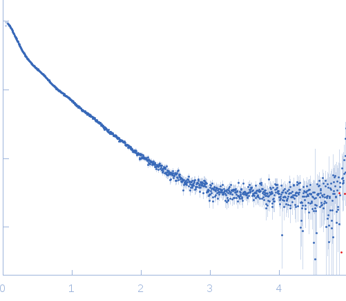
Synchrotron SAXS data from solutions of deglycosylated myelin-associated glycoprotein (Ig domains 1-5) in HEPES, pH 7.5 were collected on the BM29 camera on the storage ring ESRF (Grenoble, France) using a Pilatus 1M detector at a sample-detector distance of 2.9 m and at a wavelength of λ = 0.1 nm (I(s) vs s, where s = 4π sin θ/λ and 2θ is the scattering angle). One solute concentration of 2.13 mg/ml was measured at 20°C. 10 successive 2 second frames were collected. The data were normalized to the intensity of the transmitted beam and radially averaged; the scattering of the solvent-blank was subtracted and the curve was scaled for protein concentration.

Endo-Hf-deglycosylated full extracellular domain of Myelin-associated glycoprotein (Ig1-5).

Myelin-associated glycoprotein Ig domains 1-5 (MAG)
Mol. type   Protein
Organism   Mus musculus
Olig. state   Dimer
Mon. MW   54.0 kDa
 
UniProt   P20917 (20-508)
Sequence   FASTA
 
PDB ID   5LF5