Unique structural features of a bacterial autotransporter adhesin suggest mechanisms for interaction with host macromolecules.

Paxman JJ, Lo AW, Sullivan MJ, Panjikar S, Kuiper M, Whitten AE, Wang G, Luan CH, Moriel DG, Tan L, Peters KM, Phan MD, Gee CL, Ulett GC, Schembri MA, Heras B, Nat Commun 10(1):1967 (2019) Europe PMC

SASDC45 – Alpha domain of autotransporter protein UpaB from UPEC

Alpha domain of autotransporter protein UpaB
MWI(0) 47 kDa
MWexpected 48 kDa
VPorod 66 nm3
log I(s) 2.71×10⁻² 2.71×10⁻³ 2.71×10⁻⁴ 2.71×10⁻⁵
Alpha domain of autotransporter protein UpaB small angle scattering data  s, nm-1
ln I(s)
Alpha domain of autotransporter protein UpaB Guinier plot ln 2.71×10⁻² Rg: 2.9 nm 0 (2.9 nm)-2 s2
(sRg)2I(s)/I(0)
Alpha domain of autotransporter protein UpaB Kratky plot 1.104 0 3 sRg
p(r)
Alpha domain of autotransporter protein UpaB pair distance distribution function Rg: 3.0 nm 0 Dmax: 10.5 nm

Data validation


Fits and models


log I(s)
 s, nm-1
Alpha domain of autotransporter protein UpaB CORAL model

log I(s)
 s, nm-1
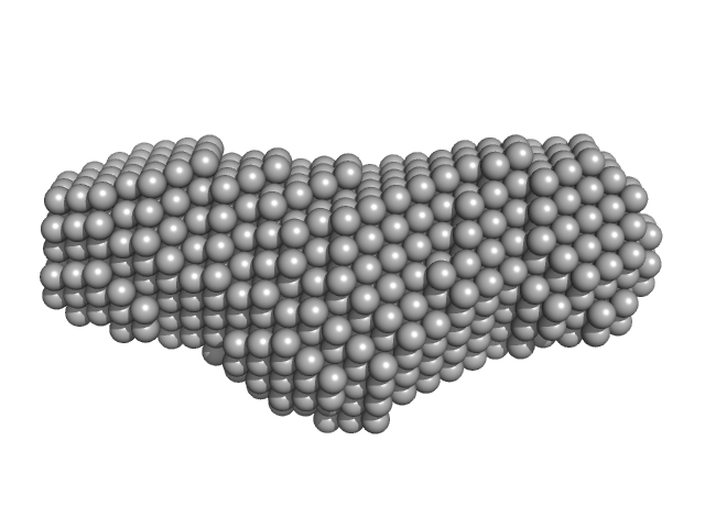
Alpha domain of autotransporter protein UpaB DAMFILT model

X-ray synchrotron radiation scattering data from solutions of the alpha domain of the autotransporter protein UpaB from uropathogenic E. coli (UPEC) in 25 mM HEPES 150mM NaCl pH 7.5 were collected on the SAXS/WAXS beam line of the Australian Synchrotron (Melbourne, Australia) using a 2D Photon counting Pilatus 1M-W pixel detector (I(s) vs s, where s = 4π sin θ/λ and 2θ is the scattering angle; λ=0.1033 nm). Thirty five successive 1 second frames were collected across solute concentrations of 0.1-2.7 mg/ml. The SAXS data displayed this entry was derived from a 0.7 mg/ml sample. The data were normalized to the intensity of the transmitted beam and radially averaged and the scattering of the solvent-blank was subtracted. The models and corresponding fits include those derived from dummy-atom modelling using DAMMIN (top) and rigid-body modelling using CORAL (bottom).

Alpha domain of autotransporter protein UpaB (Alpha-UpaB)
Mol. type   Protein
Organism   E. Coli CFT073
Olig. state   Monomer
Mon. MW   47.7 kDa
 
UniProt   A0A0H2V5A3 (37-500)
Sequence   FASTA
 
PDB ID   6BEA