The molecular basis of protein toxin HicA-dependent binding of the protein antitoxin HicB to DNA.

Winter AJ, Williams C, Isupov MN, Crocker H, Gromova M, Marsh P, Wilkinson OJ, Dillingham MS, Harmer NJ, Titball RW, Crump MP, J Biol Chem (2018) Europe PMC

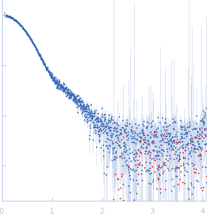
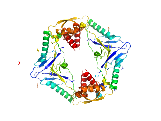
SASDD55 – Type II toxin-antitoxin system HicB family antitoxin - HicB protein - bound to addiction module toxin, HicA

Type II toxin-antitoxin system HicB family antitoxin
Addiction module toxin, HicA
MWexperimental 74 kDa
MWexpected 70 kDa
VPorod 110 nm3
log I(s) 3.63×10⁻³ 3.63×10⁻⁴ 3.63×10⁻⁵ 3.63×10⁻⁶
Type II toxin-antitoxin system HicB family antitoxin Addiction module toxin, HicA small angle scattering data  s, nm-1
ln I(s)
Type II toxin-antitoxin system HicB family antitoxin Addiction module toxin, HicA Guinier plot ln 3.64×10⁻³ Rg: 3.2 nm 0 (3.2 nm)-2 s2
(sRg)2I(s)/I(0)
Type II toxin-antitoxin system HicB family antitoxin Addiction module toxin, HicA Kratky plot 1.104 0 3 sRg
p(r)
Type II toxin-antitoxin system HicB family antitoxin Addiction module toxin, HicA pair distance distribution function Rg: 3.2 nm 0 Dmax: 11 nm

Data validation


Fits and models


log I(s)
 s, nm-1
Type II toxin-antitoxin system HicB family antitoxin Addiction module toxin, HicA PYMOL model

Synchrotron SAXS data from solutions of HicB bound to HicA in 25 mM Tris 150 mM NaCl, pH 7.5 were collected using in-line SEC-SAXS at the B21 beam line at the Diamond Light Source (Oxfordshire, UK) using a Pilatus 2M detector at a sample-detector distance of 4.0 m and at a wavelength of λ = 0.1 nm (l(s) vs s, where s = 4πsinθ/λ, and 2θ is the scattering angle). In line SEC-SAXS was performed using an Agilent 1200 HPLC system equipped with a 2.4 mL Superdex S200 column (GE Healthcare). A sample concentration of 5mg/ml was loaded onto the column at a flow rate of 0.04 mL/min. Data frames were collected at 25°C for the entire column elution with an exposure time of 3 seconds per frame through an angular s-range of 0.04-4 nm-1. The data were normalized to the intensity of the transmitted beam and radially averaged; the scattering of the solvent-blank was subtracted using data frames measured from the flow-through column buffer with no macromolecular sample present.

Analysis of the data involved ScÅtter (www.bioisis.net/). The resultant curve was fit with the crystal structure of HicB, PDB Code: 6G26.

Type II toxin-antitoxin system HicB family antitoxin (HicB)
Mol. type   Protein
Organism   Burkholderia pseudomallei
Olig. state   Tetramer
Mon. MW   15.7 kDa
 
UniProt   Q63NA5 (1-134)
Sequence   FASTA
 
Addiction module toxin, HicA (HicA)
Mol. type   Protein
Organism   Burkholderia pseudomallei
Olig. state   Monomer
Mon. MW   7.1 kDa
 
UniProt   Q63NA6
Sequence   FASTA
 
PDB ID   6G26