Molecular Fingerprints for a Novel Enzyme Family in Actinobacteria with Glucosamine Kinase Activity.

Manso JA, Nunes-Costa D, Macedo-Ribeiro S, Empadinhas N, Pereira PJB, MBio 10(3) (2019) Europe PMC

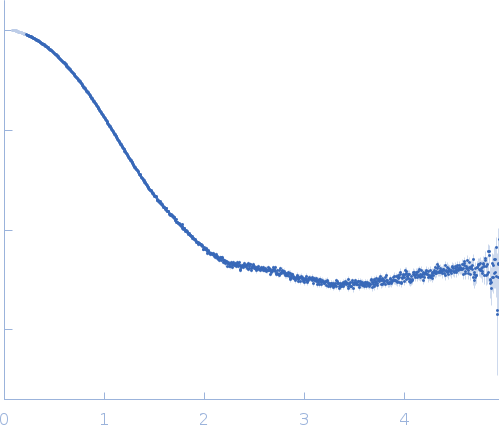
SASDEN6 – Glucosamine kinase from Streptacidiphilus jiangxiensis in presence of 1 mM ATP

Glucosamine kinase
MWI(0) 51 kDa
MWexpected 48 kDa
VPorod 73 nm3
log I(s) 4.47×101 4.47×100 4.47×10⁻¹ 4.47×10⁻²
Glucosamine kinase small angle scattering data  s, nm-1
ln I(s)
Glucosamine kinase Guinier plot ln 4.48×101 Rg: 2.5 nm 0 (2.5 nm)-2 s2
(sRg)2I(s)/I(0)
Glucosamine kinase Kratky plot 1.104 0 3 sRg
p(r)
Glucosamine kinase pair distance distribution function Rg: 2.5 nm 0 Dmax: 7.8 nm

Data validation


Fits and models


log I(s)
 s, nm-1
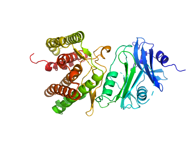
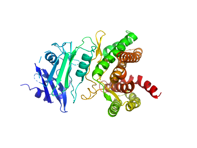
Glucosamine kinase PDB (PROTEIN DATA BANK) model

Synchrotron SAXS data from solutions of Streptacidiphilus jiangxiensis glucosamine kinase in presence of 1 mM ATP in 20 mM Tris-HCl (pH 8.0), 150 mM NaCl, 10 mM MgCl2 and 5 mM DTT were collected on the BM29 BIOSAXS beam line at the ESRF (Grenoble, France) using a Pilatus 1M detector at a sample-detector distance of 2.867 m and at a wavelength of λ = 0.0992 nm (l(s) vs s, where s = 4πsinθ/λ, and 2θ is the scattering angle; s-range = 0.035 < s < 4.944 nm-1). Solute concentrations ranging between 1.25 and 10.0 mg/ml were measured at 10°C. 10 successive 1 second frames were collected. The data were normalized to the intensity of the transmitted beam and radially averaged; the scattering of the solvent-blank was subtracted. The data were extrapolated to infinite dilution. The volume fractions for the structure above (open conformation) and below (closed conformation) were of 0.75 and 0.25 as determined by OLIGOMER, respectively.

Glucosamine kinase
Mol. type   Protein
Organism   Streptacidiphilus jiangxiensis
Olig. state   Monomer
Mon. MW   48.4 kDa
 
UniProt   A0A1H7TQR5
Sequence   FASTA
 
PDB ID   6HWJ