|
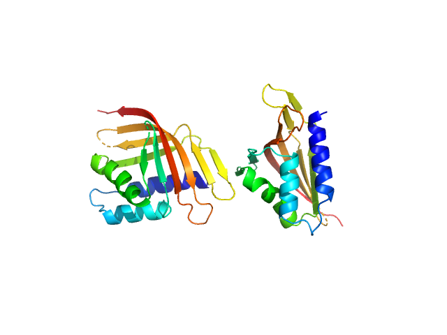
Synchrotron SAXS data from solutions of Ldb1 self association domain (L87K) in 20 mM Tris, 150 mM NaCl, 1 mM TCEP, pH 8 were collected on the SAXS/WAXS beam line at the Australian Synchrotron (Melbourne, Australia) using a Pilatus 1M detector at a sample-detector distance of 2.7 m and at a wavelength of λ = 0.103 nm (I(s) vs s, where s = 4πsinθ/λ, and 2θ is the scattering angle). One solute concentration of 0.40 mg/ml was measured at 13.6°C. 24 successive 1 second frames were collected. The data were normalized to the intensity of the transmitted beam and radially averaged; the scattering of the solvent-blank was subtracted.
A three point concentration series was collected at the Australian Synchrotron and reduced to I(s) versus s. Parameters derived from the Guinier analysis showed a small concentration dependence of systematically increasing molecular mass (M) values from I(0). The M values were consistent with a predominantly dimeric for for all samples, the data suggest there may be somewhat less than complete dimerization at the lower concentration values, and potentially some further associations at the highest concentration values. Data is provided for the sample at 0.4 mg/ml where the M value is consistent with dimer.
|
|
 s, nm-1

