Insights on the dynamic behavior of protein disulfide isomerase in the solution environment through the SAXS technique

Sanyasi C, Balakrishnan S, Chinnasamy T, Venugopalan N, Kandavelu P, Batra-Safferling R, Muthuvel S, In Silico Pharmacology 12(1) (2024) DOI

SASDH22 – Protein Disulfide Isomerase (PDI) in complex with Bovine Pancreatic Trypsin Inhibitor (BPTI)

Protein disulfide-isomerase
Pancreatic trypsin inhibitor
MWI(0) 63 kDa
MWexpected 64 kDa
VPorod 88 nm3
log I(s) 3.08×101 3.08×100 3.08×10⁻¹ 3.08×10⁻²
Protein disulfide-isomerase Pancreatic trypsin inhibitor small angle scattering data  s, nm-1
ln I(s)
Protein disulfide-isomerase Pancreatic trypsin inhibitor Guinier plot ln 3.08×101 Rg: 3.2 nm 0 (3.2 nm)-2 s2
(sRg)2I(s)/I(0)
Protein disulfide-isomerase Pancreatic trypsin inhibitor Kratky plot 1.104 0 3 sRg
p(r)
Protein disulfide-isomerase Pancreatic trypsin inhibitor pair distance distribution function Rg: 3.0 nm 0 Dmax: 8.9 nm

Data validation


Fits and models


log I(s)
 s, nm-1
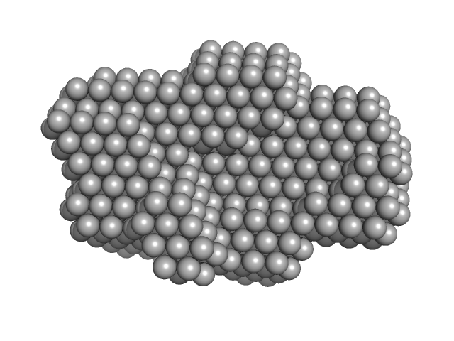
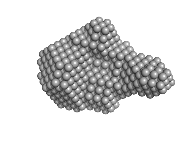
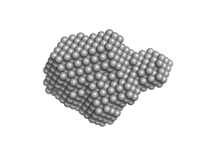
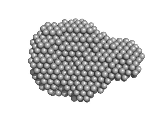
Protein disulfide-isomerase Pancreatic trypsin inhibitor DAMMIF model

log I(s)
 s, nm-1
Protein disulfide-isomerase Pancreatic trypsin inhibitor DAMMIF model

log I(s)
 s, nm-1
Protein disulfide-isomerase Pancreatic trypsin inhibitor DAMFILT model

Synchrotron SAXS data from solutions of Protein Disulfide Isomerase (PDI) in complex with Bovine Pancreatic Trypsin Inhibitor (BPTI) in 50 mM Tris-HCL, 150 mM NaCl, pH 7.5 were collected on the BioCAT 18ID beam line at the Advanced Photon Source (APS), Argonne National Laboratory (Lemont, IL, USA) using a Pilatus3 X 1M detector at a sample-detector distance of 3.5 m and at a wavelength of λ = 0.1033 nm (I(s) vs s, where s = 4πsinθ/λ, and 2θ is the scattering angle). Solute concentrations ranging between 2.5 and 10 mg/ml were measured at 10°C. Eight successive 1 second frames were collected. Using the automated data processing pipeline at the beam line, the data were normalized to the intensity of the transmitted beam and radially averaged; the scattering of the solvent-blank was subtracted. The low angle data collected at lower concentration were merged with the highest concentration high angle data to yield the final composite scattering curve. Subsequent data processing and three-dimensional modeling were performed using the ATSAS [2.8.3] package.

PDI was purified from bovine liver. Bovine Pancreatic Trypsin Inhibitor (BPTI) was purchased from SIGMA-ALDRICH in the form of lyophilized powder, dissolved with suspension medium and used directly without any further purification. CAUTION! Sample is likely aggregated.

Protein disulfide-isomerase (PDI)
Mol. type   Protein
Organism   Bos taurus
Olig. state   Monomer
Mon. MW   57.3 kDa
 
UniProt   P05307 (1-510)
Sequence   FASTA
 
Pancreatic trypsin inhibitor (BPTI)
Mol. type   Protein
Organism   Bos taurus
Olig. state   Monomer
Mon. MW   6.5 kDa
 
UniProt   P00974 (36-93)
Sequence   FASTA