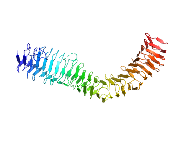
The antigen 43 structure reveals a molecular Velcro-like mechanism of autotransporter-mediated bacterial clumping.

Heras B, Totsika M, Peters KM, Paxman JJ, Gee CL, Jarrott RJ, Perugini MA, Whitten AE, Schembri MA, Proc Natl Acad Sci U S A 111(1):457-62 (2014) Europe PMC

SASDH23 – Antigen 43 alpha domain

Alpha domain of Ag43a
MWI(0) 40 kDa
MWexpected 49 kDa
VPorod 62 nm3
log I(s) 1.12×10⁻¹ 1.12×10⁻² 1.12×10⁻³ 1.12×10⁻⁴
Alpha domain of Ag43a small angle scattering data  s, nm-1
ln I(s)
Alpha domain of Ag43a Guinier plot ln 1.12×10⁻¹ Rg: 3.6 nm 0 (3.6 nm)-2 s2
(sRg)2I(s)/I(0)
Alpha domain of Ag43a Kratky plot 1.104 0 3 sRg
p(r)
Alpha domain of Ag43a pair distance distribution function Rg: 3.5 nm 0 Dmax: 12.2 nm

Data validation


Fits and models


log I(s)
 s, nm-1
Alpha domain of Ag43a DAMMIN model

log I(s)
 s, nm-1
Alpha domain of Ag43a PDB (PROTEIN DATA BANK) model

X-ray synchrotron radiation scattering data from Ag43a in 25 mM HEPES 150 mM NaCl, pH 7.0 were collected on the SAXS/WAXS beam line of the Australian Synchrotron (Melbourne, Australia) using a 2D Photon counting Pilatus 1M-W pixel detector (s = 4π sin θ/λ, where 2θ is the scattering angle). Six successive 2 second frames were collected at 3.4 mg/ml. The data were normalized to the intensity of the transmitted beam and radially averaged and the scattering of the solvent-blank was subtracted. The models and corresponding fits include those derived from dummy-atom modelling using DAMMIN, and a fit to the predicted scattering curve of the crystal structure using CRYSOL. Of note, the intensity error estimates are given as 2-standard errors, thus, the Chi values are a factor of two times lower in the log files than is actually the case.

Alpha domain of Ag43a (Ag43)
Mol. type   Protein
Organism   Escherichia coli
Olig. state   Monomer
Mon. MW   49.1 kDa
Sequence   FASTA
 
PDB ID   4KH3