Structure and substrate specificity determinants of the taurine biosynthetic enzyme cysteine sulphinic acid decarboxylase.

Mahootchi E, Raasakka A, Luan W, Muruganandam G, Loris R, Haavik J, Kursula P, J Struct Biol :107674 (2020) Europe PMC

SASDJR7 – Mouse cysteine sulphinic acid decarboxylase (CSAD)

Cysteine sulfinic acid decarboxylase
MWexperimental 110 kDa
MWexpected 117 kDa
VPorod 198 nm3
log I(s) 1.71×10⁻¹ 1.71×10⁻² 1.71×10⁻³ 1.71×10⁻⁴
Cysteine sulfinic acid decarboxylase small angle scattering data  s, nm-1
ln I(s)
Cysteine sulfinic acid decarboxylase Guinier plot ln 1.72×10⁻¹ Rg: 3.4 nm 0 (3.4 nm)-2 s2
(sRg)2I(s)/I(0)
Cysteine sulfinic acid decarboxylase Kratky plot 1.104 0 3 sRg
p(r)
Cysteine sulfinic acid decarboxylase pair distance distribution function Rg: 3.5 nm 0 Dmax: 15 nm

Data validation


Fits and models


log I(s)
 s, nm-1
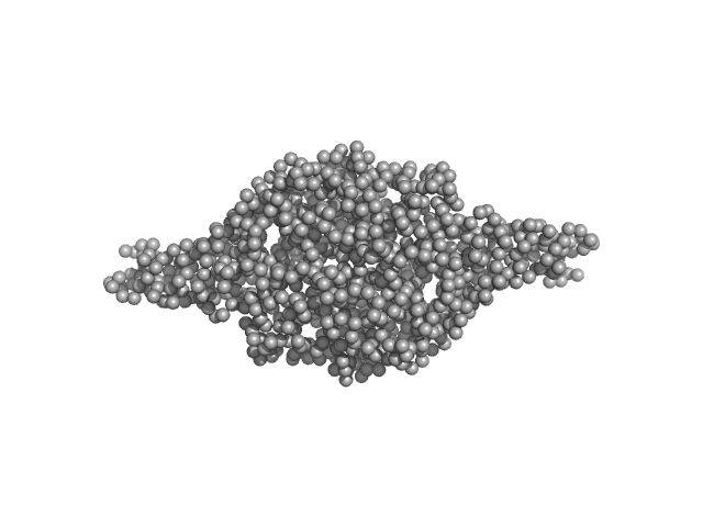
Cysteine sulfinic acid decarboxylase SREFLEX model

log I(s)
 s, nm-1
Cysteine sulfinic acid decarboxylase GASBOR model

Synchrotron SAXS data from solutions of CSAD in 20 mM HEPES, 200 mM NaCl, pH 7.5 were collected on the SWING beam line at SOLEIL (Saint-Aubin, France) using a Eiger 4M detector (I(s) vs s, where s = 4πsinθ/λ, and 2θ is the scattering angle). One solute concentration of 2.50 mg/ml was measured. The data were normalized to the intensity of the transmitted beam and radially averaged; the scattering of the solvent-blank was subtracted.

X-ray wavelength, λ = UNKNOWN; X-ray exposure time = UNKNOWN. Experimental temperature = UNKNOWN.

Cysteine sulfinic acid decarboxylase (Csad)
Mol. type   Protein
Organism   Mus musculus
Olig. state   Dimer
Mon. MW   58.4 kDa
 
UniProt   Q9DBE0 (1-493)
Sequence   FASTA
 
PDB ID   6ZEK