Elucidating Collagen Degradation Synergy between Col G and Col H from Hathewaya (Clostridium) histolytica and Identifying novel structural features in HPT and REC domains from VarS histidine kinase in V. alginolyticus

Perry Caviness, University of Arkansas PhD thesis 28030553 (2020) URL

SASDJY6 – Collagenase H C-terminal non-catalytic segments Polycystic Kidney disease domain 1 (PKD1), Polycystic Kidney disease domain 2 (PKD2) and Collagen binding domain (CBD)

Collagenase ColH (Polycystic kidney disease 1 (PKD1), Polycystic kidney disease domain 2 (PKD2) and Collagen binding domain (CBD))
MWexperimental 38 kDa
MWexpected 34 kDa
VPorod 32 nm3
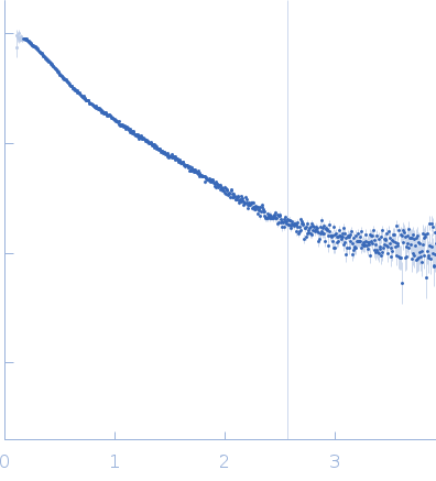
log I(s) 7.72×100 7.72×10⁻¹ 7.72×10⁻² 7.72×10⁻³
Collagenase ColH (Polycystic kidney disease 1 (PKD1), Polycystic kidney disease domain 2 (PKD2) and Collagen binding domain (CBD)) small angle scattering data  s, nm-1
ln I(s)
Collagenase ColH (Polycystic kidney disease 1 (PKD1), Polycystic kidney disease domain 2 (PKD2) and Collagen binding domain (CBD)) Guinier plot ln 7.72×100 Rg: 3.4 nm 0 (3.4 nm)-2 s2
(sRg)2I(s)/I(0)
Collagenase ColH (Polycystic kidney disease 1 (PKD1), Polycystic kidney disease domain 2 (PKD2) and Collagen binding domain (CBD)) Kratky plot 1.104 0 3 sRg
p(r)
Collagenase ColH (Polycystic kidney disease 1 (PKD1), Polycystic kidney disease domain 2 (PKD2) and Collagen binding domain (CBD)) pair distance distribution function Rg: 3.5 nm 0 Dmax: 11 nm

Data validation


Fits and models


log I(s)
 s, nm-1
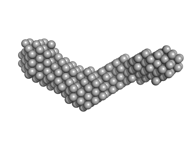
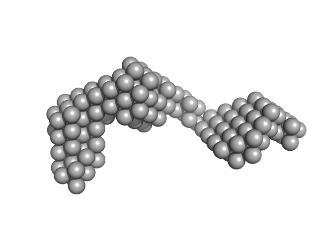
Collagenase ColH (Polycystic kidney disease 1 (PKD1), Polycystic kidney disease domain 2 (PKD2) and Collagen binding domain (CBD)) DAMFILT model

Synchrotron SAXS data from solutions of Collagenase H C-terminal non-catalytic segments Polycystic Kidney disease domain 1 (PKD1), Polycystic Kidney disease domain 2 (PKD2) and Collagen binding domain (CBD) in 10 mM HEPES, 100 mM NaCl, 0.4 mM EGTA, 2.4 mM CaCl2, pH 7.5 were collected on the 12.3.1 (SIBYLS) beam line at the Advanced Light Source (ALS) storage ring (Berkeley, CA, USA) using a Pilatus3 X 2M detector at a sample-detector distance of 1.5 m and at a wavelength of λ = 0.1127 nm (I(s) vs s, where s = 4πsinθ/λ, and 2θ is the scattering angle). Solute concentrations ranging between 1 and 3 mg/ml were measured at 10°C. 33 successive 0.300 second frames were collected. The data were normalized to the intensity of the transmitted beam and radially averaged; the scattering of the solvent-blank was subtracted. The low angle data collected at lower concentrations were extrapolated to infinite dilution and merged with the higher concentration data to yield the final composite scattering curve.

Storage temperature = UNKNOWN

Collagenase ColH (Polycystic kidney disease 1 (PKD1), Polycystic kidney disease domain 2 (PKD2) and Collagen binding domain (CBD)) (ColH)
Mol. type   Protein
Organism   Hathewaya histolytica
Olig. state   Monomer
Mon. MW   33.6 kDa
 
UniProt   Q46085 (718-1021)
Sequence   FASTA