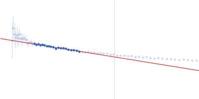
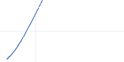
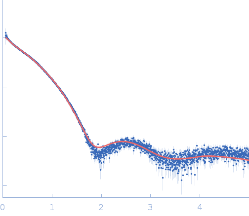
Self-assembly and conformational heterogeneity of the AXH domain of ataxin-1: an unusual example of a chameleon fold.

de Chiara C, Rees M, Menon RP, Pauwels K, Lawrence C, Konarev PV, Svergun DI, Martin SR, Chen YW, Pastore A, Biophys J 104(6):1304-13 (2013) Europe PMC

SASDL47 – Oligomeric composition of AXH Domain of Ataxin-1 (wild type and A567G, I580A mutants)

Ataxin-1
MWI(0) 45 kDa
MWexpected 14 kDa
log I(s) 3.96×101 3.96×100 3.96×10⁻¹ 3.96×10⁻²
Ataxin-1 small angle scattering data  s, nm-1
ln I(s)
Ataxin-1 Guinier plot ln 3.97×101 Rg: 5.5 nm 0 (5.5 nm)-2 s2
(sRg)2I(s)/I(0)
Ataxin-1 Kratky plot 1.104 0 3 sRg

Data validation


Fits and models


log I(s)
 s, nm-1
Ataxin-1 PDB (PROTEIN DATA BANK) model

Synchrotron SAXS data from solutions of Oligomeric composition of AXH Domain of Ataxin-1 (wild type and A567G, I580A mutants) in 20 mM Tris-HCl, pH 7 were collected on the EMBL X33 beam line at the DORIS III, DESY storage ring (Hamburg, Germany) using a Pilatus 1M-W detector at a sample-detector distance of 2.7 m and at a wavelength of λ = 0.154 nm (I(s) vs s, where s = 4πsinθ/λ, and 2θ is the scattering angle). One solute concentration of 9.30 mg/ml was measured at 10°C. Eight successive 15 second frames were collected. The data were normalized to the intensity of the transmitted beam and radially averaged; the scattering of the solvent-blank was subtracted.

OLIGOMER analysis of concentration series data, including models and fits, are made available in the full entry zip archive for the wild-type, A567G and I580A mutant variants of the protein.

Tags: X33
Ataxin-1 (AXH domain Ataxin-1)
Mol. type   Protein
Organism   Homo sapiens
Olig. state   Monomer
Mon. MW   14.0 kDa
 
UniProt   P54253 (567-689)
Sequence   FASTA
 
PDB ID   1OA8
 
PDB ID   1OA8
 
PDB ID   1OA8
 
PDB ID   1OA8