A structural basis for Staphylococcal complement subversion: X-ray structure of the complement-binding domain of Staphylococcus aureus protein Sbi in complex with ligand C3d

Clark E, Crennell S, Upadhyay A, Zozulya A, Mackay J, Svergun D, Bagby S, van den Elsen J, Molecular Immunology 48(4):452-462 (2011) DOI

SASDLF3 – Staphylococcal immunoglobulin-binding protein bound to complement component C3

Protein A
MWexperimental 33 kDa
MWexpected 28 kDa
VPorod 81 nm3
log I(s) 1.00×10⁴ 1.00×10³ 1.00×10² 1.00×101
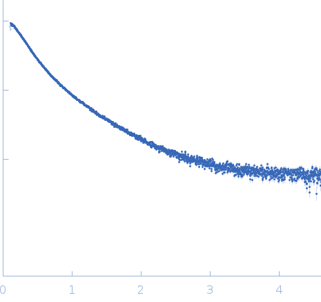
Protein A small angle scattering data  s, nm-1
ln I(s)
Protein A Guinier plot ln 1.00×10⁴ Rg: 4.6 nm 0 (4.6 nm)-2 s2
(sRg)2I(s)/I(0)
Protein A Kratky plot 1.104 0 3 sRg
p(r)
Protein A pair distance distribution function Rg: 4.7 nm 0 Dmax: 16 nm

Data validation


Fits and models


log I(s)
 s, nm-1
Protein A DAMMIN model

Synchrotron SAXS data from solutions of Staphylococcal immunoglobulin-binding protein bound to complement component C3 in 10 mM HEPES, pH 7.2, 3 mM EDTA, pH 7.2 were collected on the EMBL X33 beam line at the DORIS III, DESY storage ring (Hamburg, Germany) using a MAR 345 Image Plate detector at a sample-detector distance of 2.4 m and at a wavelength of λ = 0.15 nm (I(s) vs s, where s = 4πsinθ/λ, and 2θ is the scattering angle). Solute concentrations ranging between 2.8 and 16.7 mg/ml were measured . One 180 second frame was collected. The data were normalized to the intensity of the transmitted beam and radially averaged; the scattering of the solvent-blank was subtracted. The low angle data collected at lower concentrations were extrapolated to infinite dilution and merged with the higher concentration data to yield the final composite scattering curve.

Cell temperature = UNKNOWN. Storage temperature = UNKNOWN

Tags: X33
Protein A (Sbi-E)
Mol. type   Protein
Organism   Staphylococcus aureus
Olig. state   Monomer
Mon. MW   28.3 kDa
 
UniProt   Q70AB8 (28-266)
Sequence   FASTA