|
Synchrotron SAXS data from solutions of human nerve growth factor in 50 mM Na-phosphate, 1 mM EDTA, pH 7 were collected on the EMBL X33 beam line at DORIS III (DESY, Hamburg, Germany) using a MAR 345 Image Plate detector at a sample-detector distance of 3 m and at a wavelength of λ = 0.154 nm (I(s) vs s, where s = 4πsinθ/λ, and 2θ is the scattering angle). One solute concentration of 3.77 mg/ml was measured at 20°C. Two successive 120 second frames were collected. The data were normalized to the intensity of the transmitted beam and radially averaged; the scattering of the solvent-blank was subtracted.
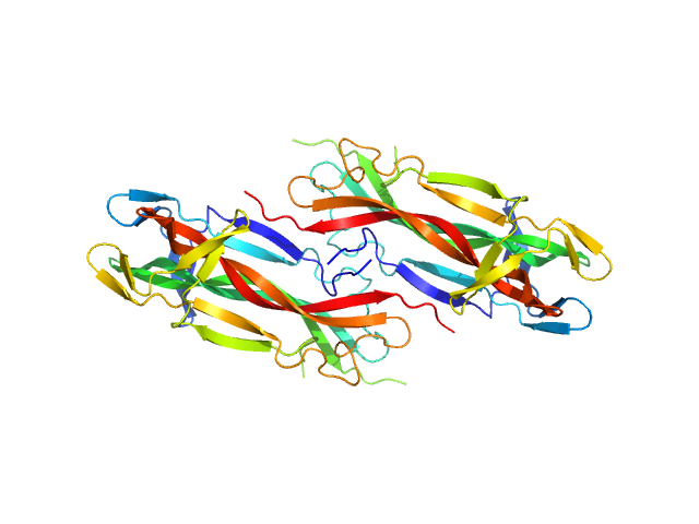
This entry displays an example SAXS profile at 3.77 mg/ml that is best described as a dimer-of-dimer equilibrium of hNGF, with the corresponding volume fractions of 62.3 and 37.7, respectively. Protein hNGF undergoes concentration dependent oligomerization. The full concentration series data (spanning 0.43-5.50 mg/ml) and the respective oligomeric analyses are made available in the full-entry zip archive.
|
|
 s, nm-1

