Structural and Functional Characterization of Rv0792c from Mycobacterium tuberculosis: Identifying Small Molecule Inhibitor against HutC Protein.

Chauhan NK, Anand A, Sharma A, Dhiman K, Gosain TP, Singh P, Singh P, Khan E, Chattopadhyay G, Kumar A, Sharma D, Ashish, Sharma TK, Singh R, Microbiol Spectr :e0197322 (2022) Europe PMC

SASDLU8 – Complex of Mtb GntR and Aptamer 5 [Rv0792c and Rv0792c_5]

Complex of GntR protein: Aptamer5
MWexperimental 86 kDa
MWexpected 78 kDa
log I(s) 1.15×10³ 1.15×10² 1.15×101 1.15×100
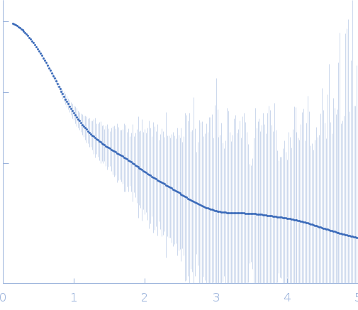
Complex of GntR protein: Aptamer5 small angle scattering data  s, nm-1
ln I(s)
Complex of GntR protein: Aptamer5 Guinier plot ln 1.16×10³ Rg: 3.4 nm 0 (3.4 nm)-2 s2
(sRg)2I(s)/I(0)
Complex of GntR protein: Aptamer5 Kratky plot 1.104 0 3 sRg
p(r)
Complex of GntR protein: Aptamer5 pair distance distribution function Rg: 3.4 nm 0 Dmax: 12 nm

Data validation


Fits and models


log I(s)
 s, nm-1
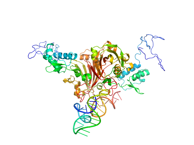
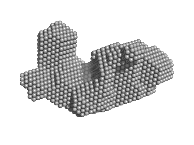
Complex of GntR protein: Aptamer5 OTHER model

log I(s)
 s, nm-1
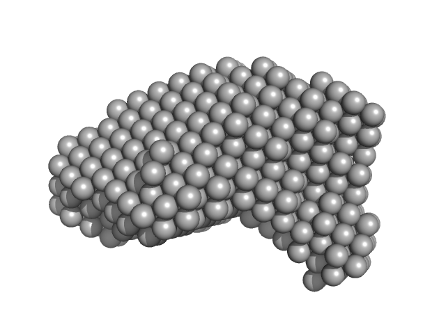
Complex of GntR protein: Aptamer5 DAMFILT model
Complex of GntR protein: Aptamer5 OTHER model
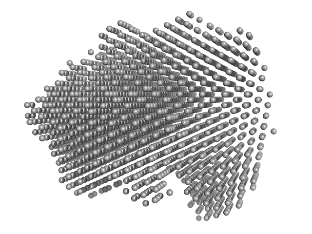
Complex of GntR protein: Aptamer5 DAMMIF model

SAXS data from solutions of Complex of Mtb GntR and Aptamer 5 [Rv0792c and Rv0792c_5] in 25 mM HEPES Buffer; 400 mM NaCl, pH 7.2 were collected at Anton Paar SAXSpace, CSIR - Institute of Microbial Technology (IMTech) using a Mythen 1K detector at a sample-detector distance of 0.3 m and at a wavelength of λ = 0.15414 nm (I(s) vs s, where s = 4πsinθ/λ, and 2θ is the scattering angle). One solute concentration of 3.00 mg/ml was measured at 10°C. Three successive 3600 second frames were collected. The data were normalized to the intensity of the transmitted beam and radially averaged; the scattering of the solvent-blank was subtracted.

Complex of GntR protein: Aptamer5
Mol. type   Other
Olig. state   Monomer
Mon. MW   78 kDa
Chemical formula