Pathogen-specific structural features of Candida albicans Ras1 activation complex: uncovering new antifungal drug targets.

Manso JA, Carabias A, Sárkány Z, de Pereda JM, Pereira PJB, Macedo-Ribeiro S, mBio :e0063823 (2023) Europe PMC

SASDM55 – GTP-binding domain of Candida albicans Ras-like protein 1

GTP-binding domain of Ras-like protein 1
MWI(0) 11 kDa
MWexpected 19 kDa
VPorod 26 nm3
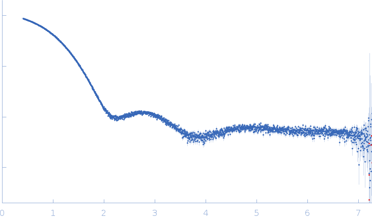
log I(s) 1.14×10² 1.14×101 1.14×100 1.14×10⁻¹
GTP-binding domain of Ras-like protein 1 small angle scattering data  s, nm-1
ln I(s)
GTP-binding domain of Ras-like protein 1 Guinier plot ln 1.14×10² Rg: 1.6 nm 0 (1.6 nm)-2 s2
(sRg)2I(s)/I(0)
GTP-binding domain of Ras-like protein 1 Kratky plot 1.104 0 3 sRg
p(r)
GTP-binding domain of Ras-like protein 1 pair distance distribution function Rg: 1.6 nm 0 Dmax: 4.1 nm

Data validation


Fits and models


log I(s)
 s, nm-1
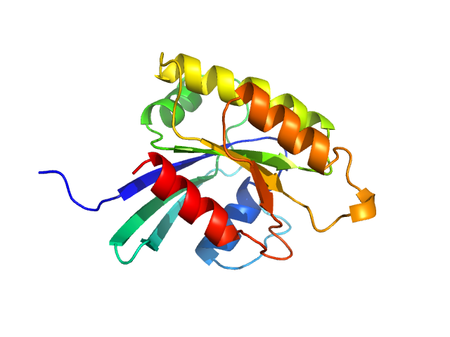
GTP-binding domain of Ras-like protein 1 MODELLER model

log I(s)
 s, nm-1
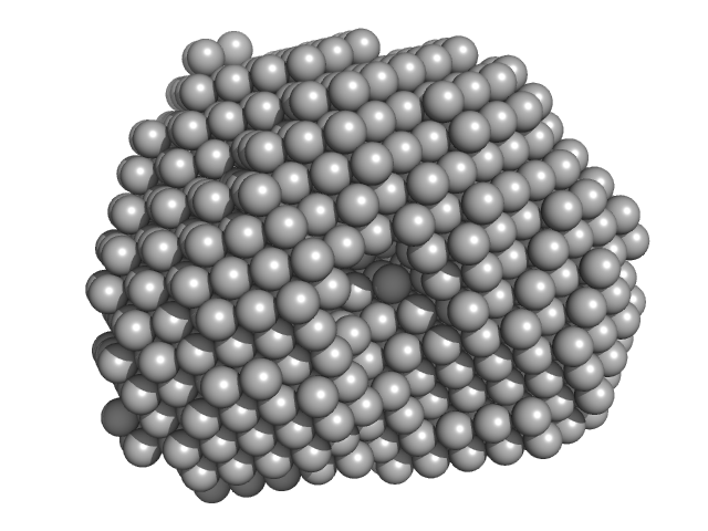
GTP-binding domain of Ras-like protein 1 DAMMIF model

Synchrotron SAXS data from solutions of the GTP-binding domain of Candida albicans Ras-like protein 1 in 20 mM Tris-HCl, 150 mM NaCl, 5% glycerol, 5 mM MgCl2, 3 mM DTT, pH 7.5 were collected on the EMBL P12 beam line at Petra-III (DESY, Hamburg, Germany) using a Pilatus 6M detector (I(s) vs s; s = 4π sin θ/λ, where 2θ is the scattering angle and λ = 0.124 nm). Different solute concentrations in the range 1.94-31 mg/ml were measured using an exposure time of 3 s (recorded as 30 x 0.1 s frames). The data were normalized to the intensity of the transmitted beam and radially averaged and the scattering from the matched solvent-blank was subtracted. The data were normalized to protein concentration and then extrapolated to infinite dilution to simulate a zero-concentration scattering curve.

Storage temperature = UNKNOWN

GTP-binding domain of Ras-like protein 1
Mol. type   Protein
Olig. state   Monomer
Mon. MW   19.0 kDa
 
UniProt   P0CY32 (2-166)
Sequence   FASTA