Structural basis underlying the synergism of NADase and SLO during group A Streptococcus infection.

Tsai WJ, Lai YH, Shi YA, Hammel M, Duff AP, Whitten AE, Wilde KL, Wu CM, Knott R, Jeng US, Kang CY, Hsu CY, Wu JL, Tsai PJ, Chiang-Ni C, Wu JJ, Lin YS, Liu CC, Senda T, Wang S, Commun Biol 6(1):124 (2023) Europe PMC

SASDM57 – NAD glycohydrolase (NADase)/Streptolysin O (SLO) complex (SAXS with additional contrast variation SANS data)

NAD glycohydrolase
Streptolysin O (T66M)
MWexperimental 101 kDa
MWexpected 110 kDa
VPorod 125 nm3
log I(s) 2.96×10² 2.96×101 2.96×100 2.96×10⁻¹
NAD glycohydrolase Streptolysin O (T66M) small angle scattering data  s, nm-1
ln I(s)
NAD glycohydrolase Streptolysin O (T66M) Guinier plot ln 2.97×10² Rg: 4.8 nm 0 (4.8 nm)-2 s2
(sRg)2I(s)/I(0)
NAD glycohydrolase Streptolysin O (T66M) Kratky plot 1.104 0 3 sRg
Dmax: 18.4 nm

Data validation


Fits and models


log I(s)
 s, nm-1
NAD glycohydrolase Streptolysin O (T66M) BILBOMD model

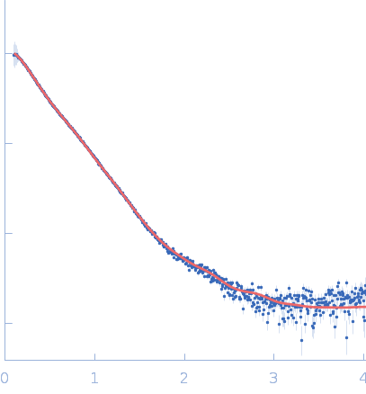
Synchrotron SAXS data from solutions of the NADase/SLO complex in phosphate buffered saline, pH 7.4 were collected on the 12.3.1 (SIBYLS) beam line at the Advanced Light Source (ALS; Berkeley, CA, USA) using a Pilatus3 X 2M detector at a sample-detector distance of 1.5 m and at a wavelength of λ = 0.103 nm (I(s) vs s, where s = 4πsinθ/λ, and 2θ is the scattering angle). In-line size-exclusion chromatography (SEC) SAS was employed. The SEC parameters were as follows: A 50.00 μl sample at 12 mg/ml was injected at a 0.50 ml/min flow rate onto a Shodex KW-800 series column at 20°C. The data were normalized to the intensity of the transmitted beam and radially averaged; the scattering of the solvent-blank was subtracted from those sample frames encompassing the SEC elution peak.

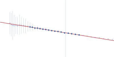
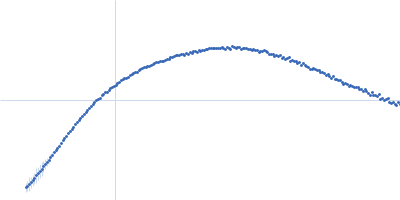
The best fit to the SAXS data is a multistate model consisting of a 9% volume fraction of the compact conformation of the complex (top) and a 53% volume fraction of the extended state (middle), in combination with a 38% volume fraction of free NADase (bottom). Small angle neutron scattering (SANS) with contrast variation data and the subsequent analysis of the contrast variation datasets are made available in the full entry zip archive.

NAD glycohydrolase (NADase)
Mol. type   Protein
Organism   Streptococcus pyogenes M1 476
Olig. state   Monomer
Mon. MW   47.2 kDa
 
UniProt   J7M2L4 (41-454)
Sequence   FASTA
 
Streptolysin O (T66M) (SLO)
Mol. type   Protein
Organism   Streptococcus pyogenes serotype M1
Olig. state   Monomer
Mon. MW   62.8 kDa
 
UniProt   P0C0I3 (32-571)
Sequence   FASTA