|
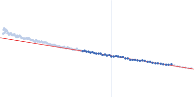
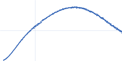
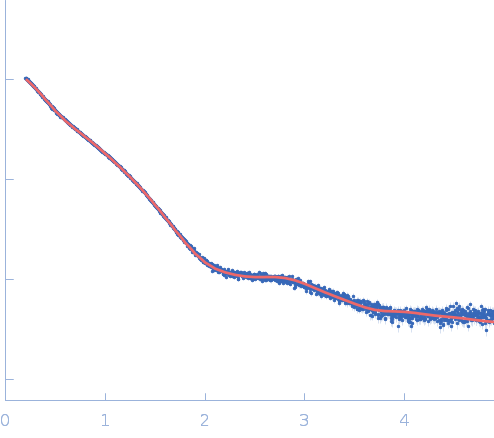
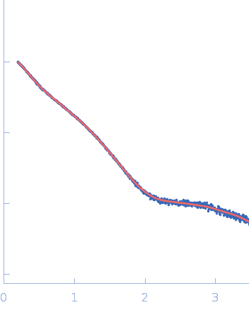
Synchrotron SAXS data from solutions of the Candida albicans Ras-like protein 1(GTP-binding domain and hypervariable region) in 20 mM Tris-HCl, 150 mM NaCl, 5% glycerol, 5 mM MgCl2, 3 mM DTT, pH 7.5 were collected on the EMBL P12 beam line at Petra-III (DESY, Hamburg, Germany) using a Pilatus 6M detector (I(s) vs s; s = 4π sin θ/λ, where 2θ is the scattering angle and λ = 0.124 nm). Different solute concentrations in the range 2.12-16.96 mg/ml were measured using an exposure time of 3 s (recorded as 30 x 0.1 s frames). The data were normalized to the intensity of the transmitted beam and radially averaged and the scattering from the matched solvent-blank was subtracted. The data were normalized to protein concentration and then extrapolated to infinite dilution to simulate a zero-concentration scattering curve. EOM analysis of the flexibility in the hypervariable region. The full EOM results are available in the full entry zip archive.
Storage temperature = UNKNOWN
|
|
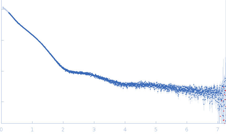 s, nm-1
 Rg, nm