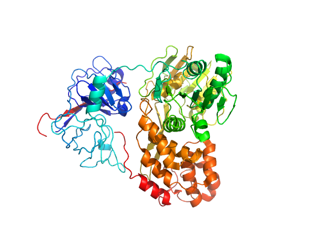
A macrocyclic HCV NS3/4A protease inhibitor interacts with protease and helicase residues in the complex with its full-length target

Schiering N, D'Arcy A, Villard F, Simic O, Kamke M, Monnet G, Hassiepen U, Svergun D, Pulfer R, Eder J, Raman P, Bodendorf U, Proceedings of the National Academy of Sciences 108(52):21052-21056 (2011) DOI

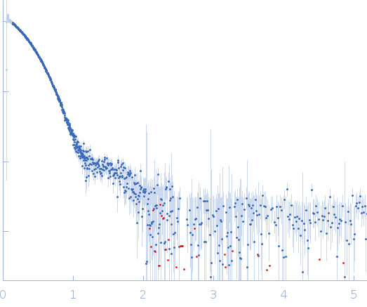
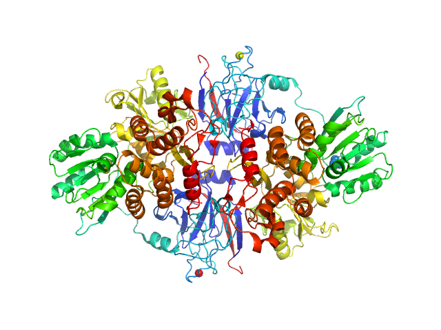
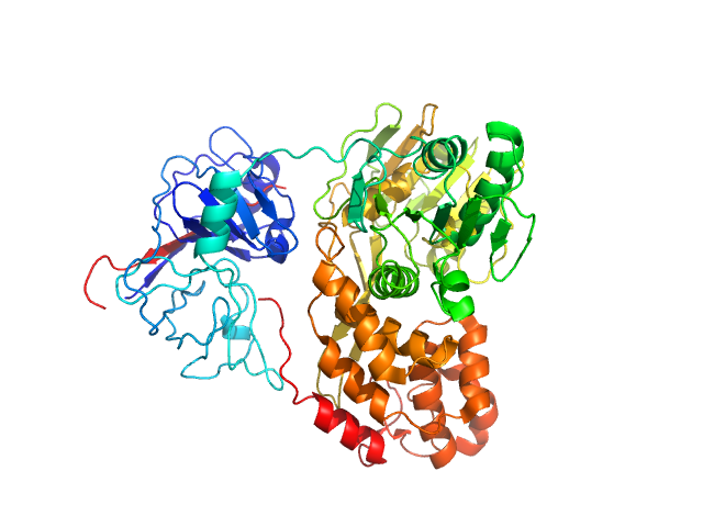
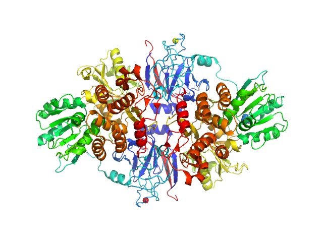
SASDML9 – Structure of bifunctional protease-helicase NS3/4A domain assembly in solution

Genome polyprotein
MWexperimental 125 kDa
MWexpected 137 kDa
log I(s) 1.70×10² 1.70×101 1.70×100 1.70×10⁻¹
Genome polyprotein small angle scattering data  s, nm-1
ln I(s)
Genome polyprotein Guinier plot ln 1.71×10² Rg: 3.9 nm 0 (3.9 nm)-2 s2
(sRg)2I(s)/I(0)
Genome polyprotein Kratky plot 1.104 0 3 sRg

Data validation


Fits and models


log I(s)
 s, nm-1
Genome polyprotein PDB (PROTEIN DATA BANK) model
Genome polyprotein PYMOL model

log I(s)
 s, nm-1
Genome polyprotein PDB (PROTEIN DATA BANK) model

log I(s)
 s, nm-1
Genome polyprotein PYMOL model

Synchrotron SAXS data from solutions of Structure of bifunctional protease-helicase NS3/4A domain assembly in solution in 25 mM Tris, 1 M NaCl, 10% glycerol, 1 mM TCEP, 0.1% β-octyl glucoside, pH 7.5 were collected on the EMBL X33 beam line at the DORIS III, DESY storage ring (Hamburg, Germany) using a Pilatus 500K detector at a sample-detector distance of 2.4 m and at a wavelength of λ = 0.15 nm (I(s) vs s, where s = 4πsinθ/λ, and 2θ is the scattering angle). One solute concentration of 7.00 mg/ml was measured. The data were normalized to the intensity of the transmitted beam and radially averaged; the scattering of the solvent-blank was subtracted.

Cell temperature = UNKNOWN. Storage temperature = UNKNOWN. Number of frames = UNKNOWN

Tags: X33
Genome polyprotein
Mol. type   Protein
Organism   Hepatitis C virus genotype 1b (isolate BK)
Olig. state   Dimer
Mon. MW   68.5 kDa
 
UniProt   P26663
Sequence   FASTA
 
PDB ID   1CU1
 
PDB ID   1CU1