Application of enhanced biophysical strategies to determine ASO/target RNA binding affinity and kinetics

Michael Lerche.

SASDN37 – ASO2 DNA in the presence of target RNA

MOE PS gapmers 3-8-3
PSCK9, 24mer ASO binding site
MWI(0) 10 kDa
MWexpected 12 kDa
VPorod 16 nm3
log I(s) 2.67×10⁻² 2.67×10⁻³ 2.67×10⁻⁴ 2.67×10⁻⁵
MOE PS gapmers 3-8-3 PSCK9, 24mer ASO binding site small angle scattering data  s, nm-1
ln I(s)
MOE PS gapmers 3-8-3 PSCK9, 24mer ASO binding site Guinier plot ln 2.68×10⁻² Rg: 2.1 nm 0 (2.1 nm)-2 s2
(sRg)2I(s)/I(0)
MOE PS gapmers 3-8-3 PSCK9, 24mer ASO binding site Kratky plot 1.104 0 3 sRg
p(r)
MOE PS gapmers 3-8-3 PSCK9, 24mer ASO binding site pair distance distribution function Rg: 2.1 nm 0 Dmax: 6.4 nm

Data validation


Fits and models


log I(s)
 s, nm-1
MOE PS gapmers 3-8-3 PSCK9, 24mer ASO binding site DAMMIF model

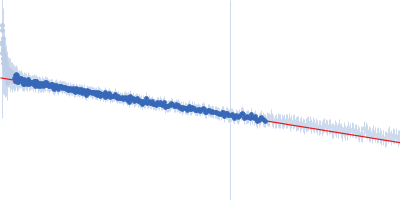
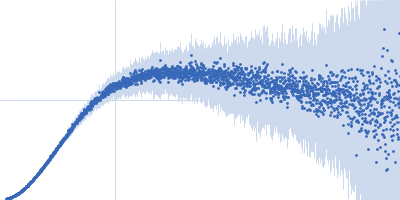
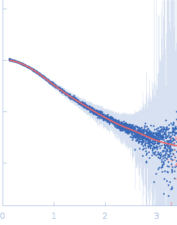
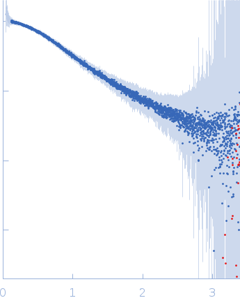
Synchrotron SAXS data from solutions of ASO2 DNA in the presence of target RNA in phosphate buffered saline, pH 7.4 were collected on the B21 beam line at the Diamond Light Source (Didcot, UK) using a Eiger 4M detector at a sample-detector distance of 0.4 m and at a wavelength of λ = 0.09537 nm (I(s) vs s, where s = 4πsinθ/λ, and 2θ is the scattering angle). 18 successive 180 second frames were collected from a sample at 15°C. The data were normalized to the intensity of the transmitted beam and radially averaged; the scattering of the solvent-blank was subtracted.

Concentration: UNKNOWN

MOE PS gapmers 3-8-3 (Aso2)
Mol. type   DNA
Olig. state   Monomer
Mon. MW   4.3 kDa
Sequence   FASTA
 
PSCK9, 24mer ASO binding site (PCSK9)
Mol. type   RNA
Olig. state   Monomer
Mon. MW   7.6 kDa
Sequence   FASTA