Solution Structure of Poly(ethylene) Glycol-Conjugated Hemoglobin Revealed by Small-Angle X-Ray Scattering: Implications for a New Oxygen Therapeutic

Svergun D, Ekström F, Vandegriff K, Malavalli A, Baker D, Nilsson C, Winslow R, Biophysical Journal 94(1):173-181 (2008) DOI

SASDNP2 – Experimental SAXS data for hemoglobin conjucted with six-seven copies of PEG dimer (Hb5)

Human hemoglobin conjugated with six-seven copies of 5-kDa PEG
MWexperimental 71 kDa
MWexpected 62 kDa
log I(s) 9.98×100 9.98×10⁻¹ 9.98×10⁻² 9.98×10⁻³
Human hemoglobin conjugated with six-seven copies of 5-kDa PEG small angle scattering data  s, nm-1
ln I(s)
Human hemoglobin conjugated with six-seven copies of 5-kDa PEG Guinier plot ln 9.98×100 Rg: 3.3 nm 0 (3.3 nm)-2 s2
(sRg)2I(s)/I(0)
Human hemoglobin conjugated with six-seven copies of 5-kDa PEG Kratky plot 1.104 0 3 sRg
p(r)
Human hemoglobin conjugated with six-seven copies of 5-kDa PEG pair distance distribution function Rg: 3.6 nm 0 Dmax: 13 nm

Data validation


Fits and models


log I(s)
 s, nm-1
Human hemoglobin conjugated with six-seven copies of 5-kDa PEG GASBOR model

log I(s)
 s, nm-1
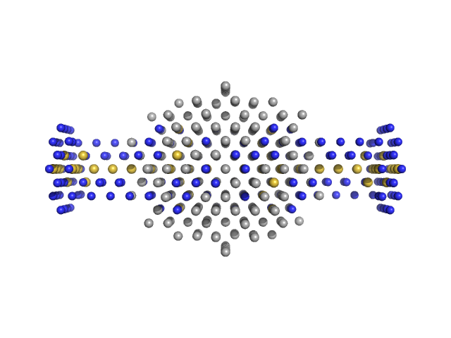
Human hemoglobin conjugated with six-seven copies of 5-kDa PEG MONSA model

Synchrotron SAXS data from solutions of Experimental SAXS data for hemoglobin conjucted with six-seven copies of PEG dimer (Hb5) in Ringer's lactate solution, pH 6.5 were collected on the EMBL X33 beam line at the DORIS III, DESY storage ring (Hamburg, Germany) using a MAR 345 Image Plate detector at a sample-detector distance of 2.5 m and at a wavelength of λ = 0.15 nm (I(s) vs s, where s = 4πsinθ/λ, and 2θ is the scattering angle). Solute concentrations ranging between 5 and 21 mg/ml were measured . Three successive 60 second frames were collected. The data were normalized to the intensity of the transmitted beam and radially averaged; the scattering of the solvent-blank was subtracted. The low angle data collected at lower concentrations were extrapolated to infinite dilution and merged with the higher concentration data to yield the final composite scattering curve.

Cell temperature = UNKNOWN. Storage temperature = UNKNOWN

Tags: X33
Human hemoglobin conjugated with six-seven copies of 5-kDa PEG (hb5)
Mol. type   Protein
Organism   Homo sapiens
Olig. state   Dimer
Mon. MW   30.8 kDa
Sequence   FASTA