Development of DNA Aptamers for Visualization of Glial Brain Tumors and Detection of Circulating Tumor Cells

Kichkailo A, Narodov A, Komarova M, Zamay T, Zamay G, Kolovskaya O, Erakhtin E, Glazyrin Y, Veprintsev D, Moryachkov R, Zabluda V, Shchugoreva I, Artyushenko P, Mironov V, Morozov D, Khorzhevskii V, Gorbushin A, Koshmanova A, Nikolaeva E, Grinev I, Voronkovskii I, Grek D, Belugin K, Volzhentsev A, Badmaev O, Luzan N, Lukyanenko K, Peters G, Lapin I, Kirichenko A, Konarev P, Morozov E, Mironov G, Gargaun A, Muharemagic D, Zamay S, Kochkina E, Dymova M, Smolyarova T, Sokolov A, Modestov A, Tokarev N, Shepelevich N, Ozerskaya A, Chanchikova N, Krat A, Zukov R, Bakhtina V, Shnyakin P, Shesternya P, Svetlichnyi V, Petrova M, Artyukhov I, Tomilin F, Berezovski M, Molecular Therapy - Nucleic Acids (2023) DOI

SASDPZ8 – Gli-233 - ssDNA aptamer specific to glioma brain tumor cells

Gli-233 - ssDNA aptamer to glioma brain tumor cells
MWexperimental 10 kDa
MWexpected 10 kDa
VPorod 13 nm3
log I(s) 2.06×101 2.06×100 2.06×10⁻¹ 2.06×10⁻²
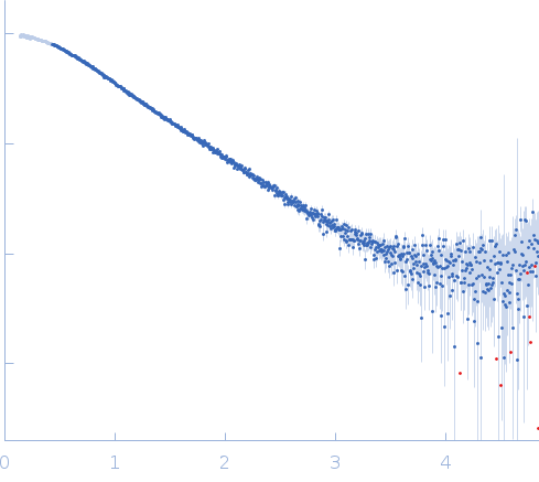
Gli-233 - ssDNA aptamer to glioma brain tumor cells small angle scattering data  s, nm-1
ln I(s)
Gli-233 - ssDNA aptamer to glioma brain tumor cells Guinier plot ln 2.06×101 Rg: 1.9 nm 0 (1.9 nm)-2 s2
(sRg)2I(s)/I(0)
Gli-233 - ssDNA aptamer to glioma brain tumor cells Kratky plot 1.104 0 3 sRg
p(r)
Gli-233 - ssDNA aptamer to glioma brain tumor cells pair distance distribution function Rg: 1.9 nm 0 Dmax: 6.8 nm

Data validation


Fits and models


log I(s)
 s, nm-1
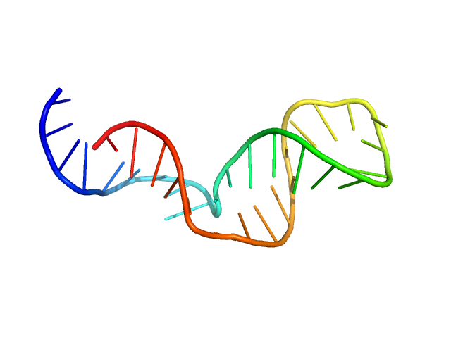
Gli-233 - ssDNA aptamer to glioma brain tumor cells GROMACS model

log I(s)
 s, nm-1
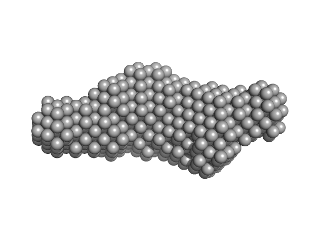
Gli-233 - ssDNA aptamer to glioma brain tumor cells DAMFILT model

Synchrotron SAXS data from Gli-233 were collected at the BM29 BioSAXS beamline at ESRF (France) by the detector Dectris Pilatus 1M. The sample-to-detector distance was 2.849 m, the wavelength λ of the X-Ray beam was 0.09919 nm. The aptamer was diluted to the concentrations of 0.8, 4.0, and 8.2 mg ml-1 in the DPBS buffer with calcium and magnesium. Additional SAXS measurements in the solution without Ca2+ and Mg2+ ions (concentrations of Gli-233 were 0.8 and 4.0 mg ml-1) were carried out to check the influence of their presence on the structure formation of the aptamer. Before SAXS, all aptamer solutions were heated to 90°C and then cooled to 4°C. The recorded range of momentum transfer was 0.1 < s < 3.5 nm-1 (is defined as s = 4π sin(θ) / λ, where 2θ is the scattering angle). Data from the three SAXS curves were extrapolated to zero concentration to exclude the concentration-dependent influence on the data.

Gli-233 - ssDNA aptamer to glioma brain tumor cells (Gli-233)
Mol. type   DNA
Organism   Artificially synthesized
Olig. state   Monomer
Mon. MW   10.2 kDa
Sequence   FASTA