Structure and Function of Gli123 Involved in Mycoplasma mobile Gliding.

Matsuike D, Tahara YO, Nonaka T, Wu HN, Hamaguchi T, Kudo H, Hayashi Y, Arai M, Miyata M, J Bacteriol :e0034022 (2023) Europe PMC

SASDQ88 – recombinant p123 expressed protein (Gli123)

p123 expressed protein
MWexperimental 273 kDa
MWexpected 248 kDa
VPorod 436 nm3
log I(s) 2.45×10⁻² 2.45×10⁻³ 2.45×10⁻⁴ 2.45×10⁻⁵
p123 expressed protein small angle scattering data  s, nm-1
ln I(s)
p123 expressed protein Guinier plot ln 2.45×10⁻² Rg: 8.2 nm 0 (8.2 nm)-2 s2
(sRg)2I(s)/I(0)
p123 expressed protein Kratky plot 1.104 0 3 sRg
p(r)
p123 expressed protein pair distance distribution function Rg: 9 nm 0 Dmax: 31.6 nm

Data validation


Fits and models


log I(s)
 s, nm-1
p123 expressed protein DAMMIN model

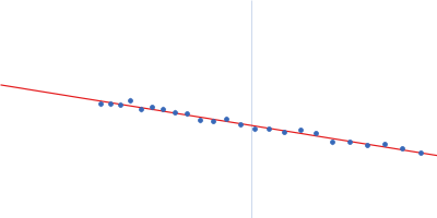
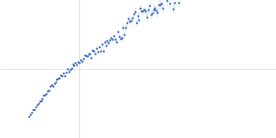
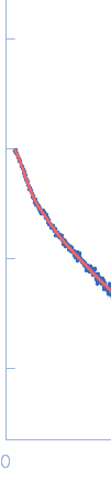
Synchrotron SAXS data from solutions of recombinant Gli123 in 100 mM ammonium acetate, pH 6.8 were collected on the BL-10C beam line at the Photon Factory (PF), High Energy Accelerator Research Organization (KEK; Tsukuba, Japan) using a Pilatus3 2M detector at a sample-detector distance of 2.1 m and at a wavelength of λ = 0.1488 nm (I(s) vs s, where s = 4πsinθ/λ, and 2θ is the scattering angle). In-line size-exclusion chromatography (SEC) SAS was employed. The SEC parameters were as follows: A 100.00 μl sample at 5.3 mg/ml was injected at a 0.10 ml/min flow rate onto a GE Superdex 200 Increase 10/300 column at 25°C. 852 successive 10 second frames were collected through the entire SEC elution. The data were normalized to the intensity of the transmitted beam and radially averaged; the scattering of the solvent-blank was subtracted.

The protein was recombinantly expressed in Escherichia coli.

p123 expressed protein (Gli123)
Mol. type   Protein
Organism   Mycoplasma mobile (strain ATCC 43663 / 163K / NCTC 11711)
Olig. state   Dimer
Mon. MW   123.9 kDa
 
UniProt   F8WJV6 (19-1128)
Sequence   FASTA