Structural analysis of the housecleaning nucleoside triphosphate pyrophosphohydrolase MazG from Mycobacterium tuberculosis

Wang S, Gao B, Chen A, Zhang Z, Wang S, Lv L, Zhao G, Li J, Frontiers in Microbiology 14 (2023) DOI

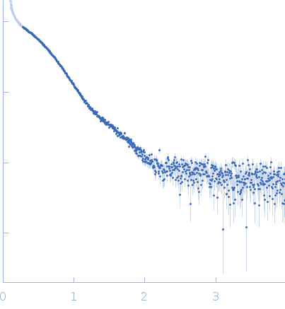
SASDQE5 – Nucleoside triphosphate pyrophosphohydrolase (1–185)

Nucleoside triphosphate pyrophosphohydrolase
MWexperimental 42 kDa
MWexpected 41 kDa
VPorod 64 nm3
log I(s) 7.28×101 7.28×100 7.28×10⁻¹ 7.28×10⁻²
Nucleoside triphosphate pyrophosphohydrolase small angle scattering data  s, nm-1
ln I(s)
Nucleoside triphosphate pyrophosphohydrolase Guinier plot ln 7.29×101 Rg: 2.7 nm 0 (2.7 nm)-2 s2
(sRg)2I(s)/I(0)
Nucleoside triphosphate pyrophosphohydrolase Kratky plot 1.104 0 3 sRg
p(r)
Nucleoside triphosphate pyrophosphohydrolase pair distance distribution function Rg: 2.6 nm 0 Dmax: 8.4 nm

Data validation


Fits and models


log I(s)
 s, nm-1
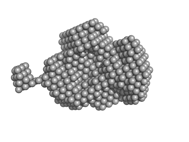
Nucleoside triphosphate pyrophosphohydrolase DAMMIF model

Synchrotron SAXS data from solutions of nucleoside triphosphate pyrophosphohydrolase (mazG) in 20 mM Tris-HCl, 100 mM NaCl, pH 7.5 were collected on the BL19U2 beam line at the Shanghai Synchrotron Radiation Facility (SSRF; Shanghai, China) using a Pilatus 1M detector at a sample-detector distance of 2.6 m and at a wavelength of λ = 0.124 nm (I(s) vs s, where s = 4πsinθ/λ, and 2θ is the scattering angle). Solute concentrations ranging between 1 and 2 mg/ml were measured at 10°C. 20 successive 1 second frames were collected. The data were normalized to the intensity of the transmitted beam and radially averaged; the scattering of the solvent-blank was subtracted.

CAUTION: Possibly aggregated.

Nucleoside triphosphate pyrophosphohydrolase (mazG)
Mol. type   Protein
Organism   Mycobacterium tuberculosis (strain ATCC 25618 / H37Rv)
Olig. state   Dimer
Mon. MW   20.3 kDa
 
UniProt   P96379 (1-185)
Sequence   FASTA