Structural insights into the multifunctionality of rabies virus P3 protein.

Sethi A, Rawlinson SM, Dubey A, Ang CS, Choi YH, Yan F, Okada K, Rozario AM, Brice AM, Ito N, Williamson NA, Hatters DM, Bell TDM, Arthanari H, Moseley GW, Gooley PR, Proc Natl Acad Sci U S A 120(14):e2217066120 (2023) Europe PMC

SASDQG4 – Rabies virus Nishigahara strain Phosphoprotein Isoform 3 (P3)

Isoform P3 of Phosphoprotein
MWexperimental 50 kDa
MWexpected 55 kDa
VPorod 108 nm3
log I(s) 3.76×10⁻³ 3.76×10⁻⁴ 3.76×10⁻⁵ 3.76×10⁻⁶
Isoform P3 of Phosphoprotein small angle scattering data  s, nm-1
ln I(s)
Isoform P3 of Phosphoprotein Guinier plot ln 3.77×10⁻³ Rg: 3.7 nm 0 (3.7 nm)-2 s2
(sRg)2I(s)/I(0)
Isoform P3 of Phosphoprotein Kratky plot 1.104 0 3 sRg
Dmax: 16.5 nm

Data validation


Fits and models


log I(s)
 s, nm-1
Rabies virus Nishigahara strain Phosphoprotein Isoform 3 (P3) Rg histogram Rg, nm
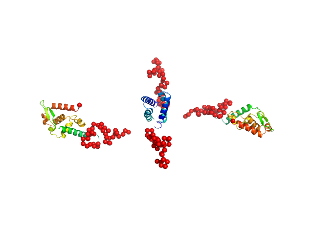
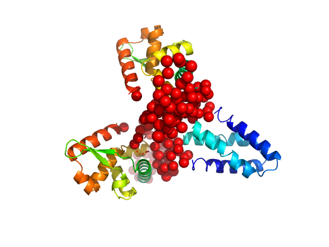
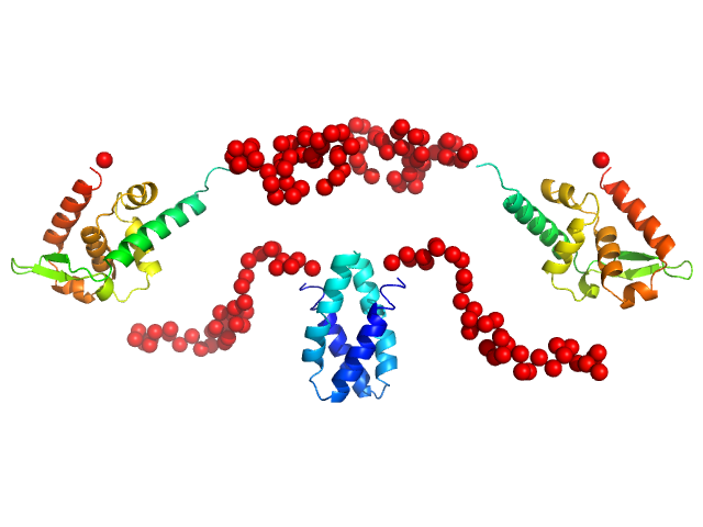
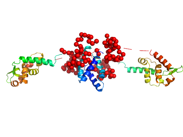
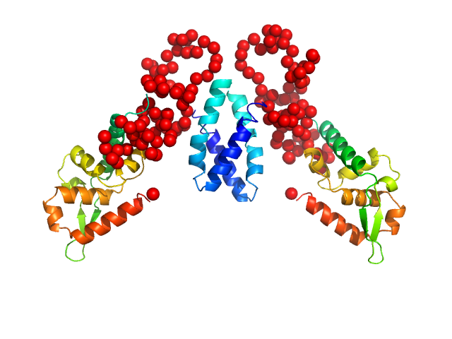
Isoform P3 of Phosphoprotein EOM/RANCH model

Synchrotron SAXS data from solutions of Rabies virus Nishigahara strain Phosphoprotein Isoform 3 (P3) in 25 mM HEPES, 150 mM NaCl, 1 mM TCEP, pH 7.4 were collected on the SAXS/WAXS beam line at the Australian Synchrotron storage ring (Melbourne, Australia) using a Pilatus3 S 2M detector at a sample-detector distance of 3.3 m and at a wavelength of λ = 0.103 nm (I(s) vs s, where s = 4πsinθ/λ, and 2θ is the scattering angle). One solute concentration of 5.00 mg/ml was measured at 22°C. One 1 second frame was collected. The data were normalized to the intensity of the transmitted beam and radially averaged; the scattering of the solvent-blank was subtracted.

Isoform P3 of Phosphoprotein (Nish P3)
Mol. type   Protein
Organism   Rabies virus (strain Nishigahara RCEH)
Olig. state   Dimer
Mon. MW   27.6 kDa
 
UniProt   Q9IPJ8-3 (1-244)
Sequence   FASTA