The structure of the second CysD domain of MUC2 and role in mucin organization by transglutaminase-based cross-linking.

Recktenwald CV, Karlsson G, Garcia-Bonete MJ, Katona G, Jensen M, Lymer R, Bäckström M, Johansson MEV, Hansson GC, Trillo-Muyo S, Cell Rep 43(5):114207 (2024) Europe PMC

SASDSG5 – Human MUC2 mucin CysD2 domain

Mucin-2 (CysD2 domain)
MWexperimental 18 kDa
MWexpected 16 kDa
VPorod 31 nm3
log I(s) 1.00×10³ 1.00×10² 1.00×101 1.00×100
Mucin-2 (CysD2 domain) small angle scattering data  s, nm-1
ln I(s)
Mucin-2 (CysD2 domain) Guinier plot ln 1.01×10³ Rg: 2.5 nm 0 (2.5 nm)-2 s2
(sRg)2I(s)/I(0)
Mucin-2 (CysD2 domain) Kratky plot 1.104 0 3 sRg
p(r)
Mucin-2 (CysD2 domain) pair distance distribution function Rg: 2.6 nm 0 Dmax: 9.3 nm

Data validation


There are no models related to this curve.

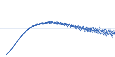
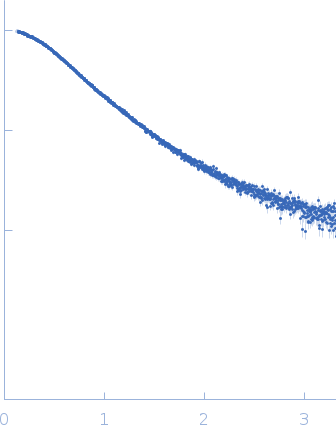
Synchrotron SAXS data from solutions of the CysD2 domain from human mucin in 20 mM sodium phosphate, 70 mM NaCl, pH 7.2 were collected on the EMBL P12 beam line at PETRA III (DESY, Hamburg, Germany) using a Pilatus 6M detector at a sample-detector distance of 3 m and at a wavelength of λ = 0.124 nm (I(s) vs s, where s = 4πsinθ/λ, and 2θ is the scattering angle). In-line size-exclusion chromatography (SEC) SAS was employed. The SEC parameters were as follows: A 50.00 μl sample at 1 mg/ml was injected at a 0.40 ml/min flow rate onto a GE Superdex 75 Increase 5/150 column at 20°C. One 0.995 second frame was collected. The data were normalized to the intensity of the transmitted beam and radially averaged; the scattering of the solvent-blank was subtracted.

Mucin-2 (CysD2 domain) (MUC2-CysD2)
Mol. type   Protein
Organism   Homo sapiens
Olig. state   Monomer
Mon. MW   16.1 kDa
 
UniProt   Q02817 (1781-1892)
Sequence   FASTA