20-kDa accessory protein (P20) from Bacillus thuringiensis subsp. israelensis ISPC-12: Purification, characterization, solution scattering and structural analysis

Kinkar O, Singh R, Prashar A, Kumar A, Hire R, Makde R, International Journal of Biological Macromolecules :127985 (2023) DOI

SASDSX8 – Solution scattering studies on 20 kDa accessory protein (P20) from Bacillus thuringiensis subsp. israelensis (ISPC-12)

20 kDa accessory protein
MWexperimental 42 kDa
MWexpected 42 kDa
VPorod 67 nm3
log I(s) 8.17×10⁻¹ 8.17×10⁻² 8.17×10⁻³ 8.17×10⁻⁴
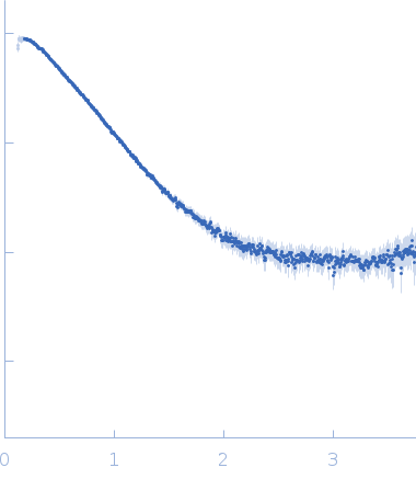
20 kDa accessory protein small angle scattering data  s, nm-1
ln I(s)
20 kDa accessory protein Guinier plot ln 8.18×10⁻¹ Rg: 3.0 nm 0 (3.0 nm)-2 s2
(sRg)2I(s)/I(0)
20 kDa accessory protein Kratky plot 1.104 0 3 sRg
p(r)
20 kDa accessory protein pair distance distribution function Rg: 2.9 nm 0 Dmax: 8.6 nm

Data validation


Fits and models


log I(s)
 s, nm-1
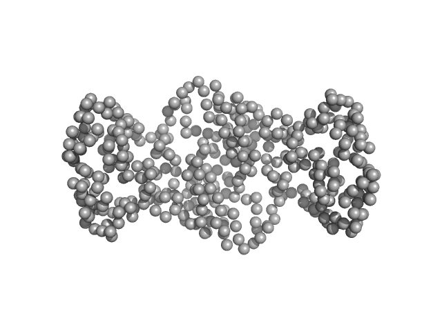
20 kDa accessory protein GASBOR model

Synchrotron SAXS data from solutions of 20 kDa accessory protein (P20) from Bacillus thuringiensis in 10 mM Tris, 100 mM NaCl, pH 8 were collected on the BL-18 beam line at INDUS-2 (Indore, India) using a MAR 345 Image Plate detector at a sample-detector distance of 2.2 m and at a wavelength of λ = 0.10332 nm (I(s) vs s, where s = 4πsinθ/λ, and 2θ is the scattering angle). One solute concentration of 5.20 mg/ml was measured at 25°C. The data were normalized to the intensity of the transmitted beam and radially averaged; the scattering of the solvent-blank was subtracted.

The P20 protein was purified by size exclusion chromatography prior to the SAXS measurement. X-ray exposure time = UNKNOWN.

20 kDa accessory protein (P20)
Mol. type   Protein
Organism   Bacillus thuringiensis serovar israelensis
Olig. state   Dimer
Mon. MW   20.9 kDa
 
UniProt   Q7AL75 (2-182)
Sequence   FASTA