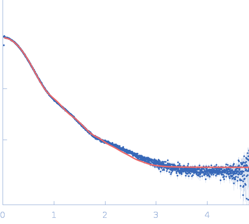
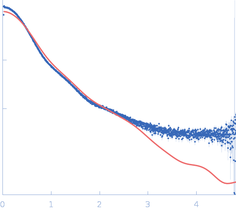
Trypanosoma brucei Invariant Surface Glycoprotein 75 Is an Immunoglobulin Fc Receptor Inhibiting Complement Activation and Antibody-Mediated Cellular Phagocytosis.

Mikkelsen JH, Stødkilde K, Jensen MP, Hansen AG, Wu Q, Lorentzen J, Graversen JH, Andersen GR, Fenton RA, Etzerodt A, Thiel S, Andersen CBF, J Immunol (2024) Europe PMC

SASDTP5 – Invariant surface glycoprotein 75 (N134A) amino acids 29-468

Invariant surface glycoprotein (N134A)
MWexperimental 54 kDa
MWexpected 49 kDa
VPorod 127 nm3
log I(s) 3.87×10⁴ 3.87×10³ 3.87×10² 3.87×101
Invariant surface glycoprotein (N134A) small angle scattering data  s, nm-1
ln I(s)
Invariant surface glycoprotein (N134A) Guinier plot ln 3.88×10⁴ Rg: 3.6 nm 0 (3.6 nm)-2 s2
(sRg)2I(s)/I(0)
Invariant surface glycoprotein (N134A) Kratky plot 1.104 0 3 sRg
p(r)
Invariant surface glycoprotein (N134A) pair distance distribution function Rg: 3.7 nm 0 Dmax: 15 nm

Data validation


Fits and models


log I(s)
 s, nm-1
Invariant surface glycoprotein (N134A) CORAL model

log I(s)
 s, nm-1
Invariant surface glycoprotein (N134A) CORAL model

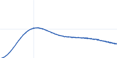
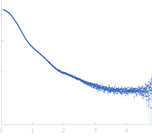
Synchrotron SAXS data from solutions of invariant surface glycoprotein 75 (N134A) in 20 mM Tris-HCl, 75 mM KCl, pH 7.6 were collected on the EMBL P12 beam line at PETRA III (DESY; Hamburg, Germany) using a Pilatus 2M detector at a sample-detector distance of 3 m and at a wavelength of λ = 0.123987 nm (I(s) vs s, where s = 4πsinθ/λ, and 2θ is the scattering angle). One solute concentration of 9.50 mg/ml was measured at 20.2°C. 18 successive 0.045 second frames were collected. The data were normalized to the intensity of the transmitted beam and radially averaged; the scattering of the solvent-blank was subtracted.

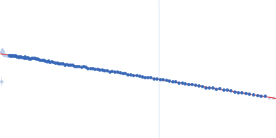
AlphaFold Protein Structure Database entry (full length monomer): https://alphafold.ebi.ac.uk/entry/Q26769

Invariant surface glycoprotein (N134A) (ISG75)
Mol. type   Protein
Organism   Trypanosoma brucei
Olig. state   Monomer
Mon. MW   49.1 kDa
 
UniProt   Q26769 (29-468)
Sequence   FASTA