Biophysical analysis of the membrane-proximal Venus Flytrap domain of ESAG4 receptor-like adenylate cyclase from Trypanosoma brucei.

Alves DO, Geens R, da Silva Arruda HR, Jennen L, Corthaut S, Wuyts E, de Andrade GC, Prosdocimi F, Cordeiro Y, Pires JR, Vieira LR, de Oliveira GAP, Sterckx YG, Salmon D, Mol Biochem Parasitol 260:111653 (2024) Europe PMC

SASDU78 – Trypanosoma brucei ESAG4 membrane-proximal Venus Fly Trap domain 2 (VFT2)

adenylate cyclase
MWexperimental 43 kDa
MWexpected 43 kDa
VPorod 76 nm3
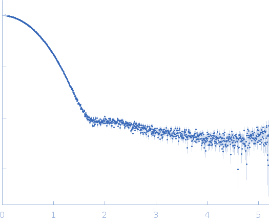
log I(s) 6.82×101 6.82×100 6.82×10⁻¹ 6.82×10⁻²
adenylate cyclase small angle scattering data  s, nm-1
ln I(s)
adenylate cyclase Guinier plot ln 6.83×101 Rg: 2.3 nm 0 (2.3 nm)-2 s2
(sRg)2I(s)/I(0)
adenylate cyclase Kratky plot 1.104 0 3 sRg
p(r)
adenylate cyclase pair distance distribution function Rg: 2.3 nm 0 Dmax: 7.6 nm

Data validation


Fits and models


log I(s)
 s, nm-1
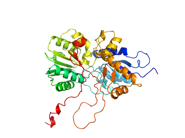
adenylate cyclase ALPHAFOLD model

log I(s)
 s, nm-1

Synchrotron SAXS data from solutions of ESAG4 venus fly trap domain 2 in 50 mM Tris-HCl, 500 mM NaCl, pH 8 were collected on the BM29 beam line at the ESRF (Grenoble, France) using a Pilatus 1M detector at a sample-detector distance of 3 m and at a wavelength of λ = 0.099 nm (I(s) vs s, where s = 4πsinθ/λ, and 2θ is the scattering angle). In-line size-exclusion chromatography (SEC) SAS was employed. The SEC parameters were as follows: A 50.00 μl sample at 9.9 mg/ml was injected at a 0.30 ml/min flow rate onto a column at 20°C. 500 successive 2 second frames were collected. The data were normalized to the intensity of the transmitted beam and radially averaged; the scattering of the solvent-blank was subtracted.

SEC column: UNKNOWN.

adenylate cyclase (VFT2)
Mol. type   Protein
Organism   Trypanosoma brucei
Olig. state   Monomer
Mon. MW   42.8 kDa
 
UniProt   Q8WPR6 (480-859)
Sequence   FASTA