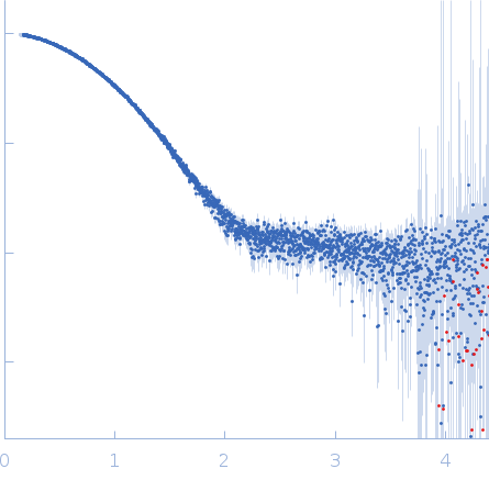
The Legionella collagen-like protein employs a distinct binding mechanism for the recognition of host glycosaminoglycans.

Rehman S, Antonovic AK, McIntire IE, Zheng H, Cleaver L, Baczynska M, Adams CO, Portlock T, Richardson K, Shaw R, Oregioni A, Mastroianni G, Whittaker SB, Kelly G, Lorenz CD, Fornili A, Cianciotto NP, Garnett JA, Nat Commun 15(1):4912 (2024) Europe PMC

SASDUJ7 – Lcl C-terminal domain R477A mutant - monomer

HbP1 (R477A)
MWexperimental 19 kDa
MWexpected 18 kDa
VPorod 33 nm3
log I(s) 7.50×10⁻² 7.50×10⁻³ 7.50×10⁻⁴ 7.50×10⁻⁵
HbP1 (R477A) small angle scattering data  s, nm-1
ln I(s)
HbP1 (R477A) Guinier plot ln 7.51×10⁻² Rg: 1.9 nm 0 (1.9 nm)-2 s2
(sRg)2I(s)/I(0)
HbP1 (R477A) Kratky plot 1.104 0 3 sRg
p(r)
HbP1 (R477A) pair distance distribution function Rg: 1.9 nm 0 Dmax: 6.4 nm

Data validation


There are no models related to this curve.

Synchrotron SAXS data from solutions of Lcl C-terminal domain R477A mutant - monomer in 20 mM Tris–HCl pH 8.0, 200 mM NaCl, 5 mM EDTA were collected on the B21 beam line at the Diamond Light Source storage ring (Didcot, UK) using a Pilatus 2M detector at a wavelength of λ = 0.1 nm (I(s) vs s, where s = 4πsinθ/λ, and 2θ is the scattering angle). One solute concentration of 5.00 mg/ml was measured at 25°C. 
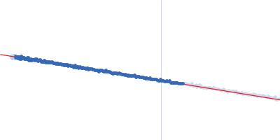
The data were normalized to the intensity of the transmitted beam and radially averaged; the scattering of the solvent-blank was subtracted.

Storage temperature = UNKNOWN. Sample detector distance = UNKNOWN. X-ray Exposure time = UNKNOWN. Number of frames = UNKNOWN
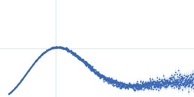
HbP1 (R477A) (Lcl-CTD R77A mon)
Mol. type   Protein
Organism   Legionella pneumophila
Olig. state   Monomer
Mon. MW   18.5 kDa
 
UniProt   E7BLH6 (387-536)
Sequence   FASTA