High Density of N- and O-Glycosylation Shields and Defines the Structural Dynamics of the Intrinsically Disordered Ectodomain of Receptor-type Protein Tyrosine Phosphatase Alpha

Chien Y, Wang Y, Sridharan D, Kuo C, Chien C, Uchihashi T, Kato K, Angata T, Meng T, Hsu S, Khoo K, JACS Au (2023) DOI

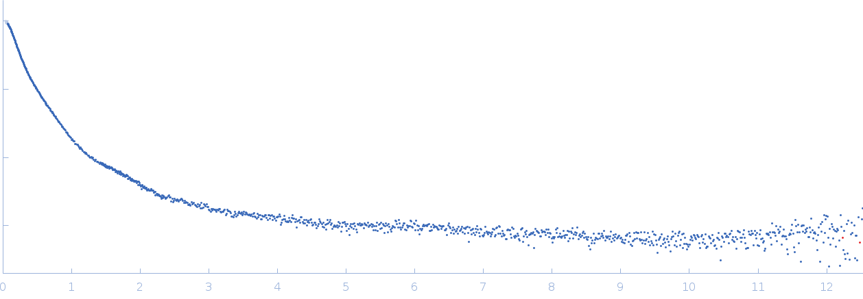
SASDV53 – Fc-fused PTPRA ECD in PBS buffer

Receptor-type tyrosine-protein phosphatase alpha
MWexperimental 271 kDa
MWexpected 150 kDa
log I(s) 5.47×10⁻¹ 5.47×10⁻² 5.47×10⁻³ 5.47×10⁻⁴
Receptor-type tyrosine-protein phosphatase alpha small angle scattering data  s, nm-1
ln I(s)
Receptor-type tyrosine-protein phosphatase alpha Guinier plot ln 5.48×10⁻¹ Rg: 8.3 nm 0 (8.3 nm)-2 s2
(sRg)2I(s)/I(0)
Receptor-type tyrosine-protein phosphatase alpha Kratky plot 1.104 0 3 sRg
Dmax: 35.3 nm

Data validation


Fits and models


log I(s)
 s, nm-1
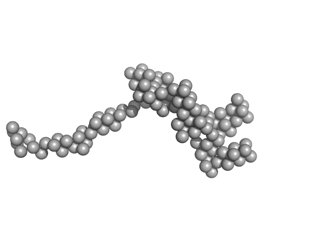
Receptor-type tyrosine-protein phosphatase alpha DAMMIF model

Synchrotron SAXS data from solutions of Fc-fused PTPRA ECD in PBS buffer in 10 mM phosphate, 137 mM NaCl, 2.7 mM KCl, pH 7.4 were collected on the 13A beam line at the Taiwan Photon Source, NSRRC storage ring (Hsinchu, Taiwan) using a Eiger X 9M detector at a sample-detector distance of 9 m and at a wavelength of λ = 0.08265 nm (I(s) vs s, where s = 4πsinθ/λ, and 2θ is the scattering angle). One solute concentration of 10.00 mg/ml was measured at 15°C. Three successive 2 second frames were collected. The data were normalized to the intensity of the transmitted beam and radially averaged; the scattering of the solvent-blank was subtracted.

Receptor-type tyrosine-protein phosphatase alpha (PTPRA)
Mol. type   Protein
Organism   Homo sapiens
Olig. state   Dimer
Mon. MW   75 kDa
Sequence   FASTA