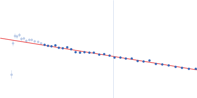
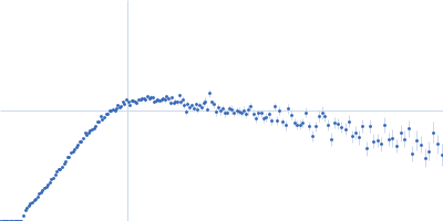
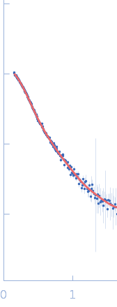
DNMT3B

CHAO-CHENG CHO.

SASDVJ9 – Human DNA methyltransferase 3 beta DNMT3B (215-853) and DNA methyltransferase 3-like DNMT3L (178-379) in complex with histone H3

Histone H3.3
DNA methyltransferase 3 beta (215-853)
MWexperimental 201 kDa
MWexpected 154 kDa
VPorod 275 nm3
log I(s) 1.68×10⁻¹ 1.68×10⁻² 1.68×10⁻³ 1.68×10⁻⁴
Histone H3.3 DNA methyltransferase 3 beta (215-853) small angle scattering data  s, nm-1
ln I(s)
Histone H3.3 DNA methyltransferase 3 beta (215-853) Guinier plot ln 1.69×10⁻¹ Rg: 5.1 nm 0 (5.1 nm)-2 s2
(sRg)2I(s)/I(0)
Histone H3.3 DNA methyltransferase 3 beta (215-853) Kratky plot 1.104 0 3 sRg
p(r)
Histone H3.3 DNA methyltransferase 3 beta (215-853) pair distance distribution function Rg: 5.2 nm 0 Dmax: 18.9 nm

Data validation


Fits and models


log I(s)
 s, nm-1
Histone H3.3 DNA methyltransferase 3 beta (215-853) GASBOR model

Synchrotron SAXS data from solutions of Human DNA methyltransferase 3 beta DNMT3B (215-853) and DNA methyltransferase 3-like DNMT3L (178-379) in complex with histone H3 in 20 mM Tris, 300 mM NaCl, 1 mM TCEP, 5% glycerol, pH 8 were collected on the 13A beam line at the Taiwan Photon Source, NSRRC storage ring (Hsinchu, Taiwan) using a Eiger X 1M and Eiger X 9M detector at a sample-detector distance of 2.5 m and at a wavelength of λ = 0.0827 nm (I(s) vs s, where s = 4πsinθ/λ, and 2θ is the scattering angle). One solute concentration of 5.00 mg/ml was measured at 10°C. 199 successive 0.200 second frames were collected. The data were normalized to the intensity of the transmitted beam and radially averaged; the scattering of the solvent-blank was subtracted.
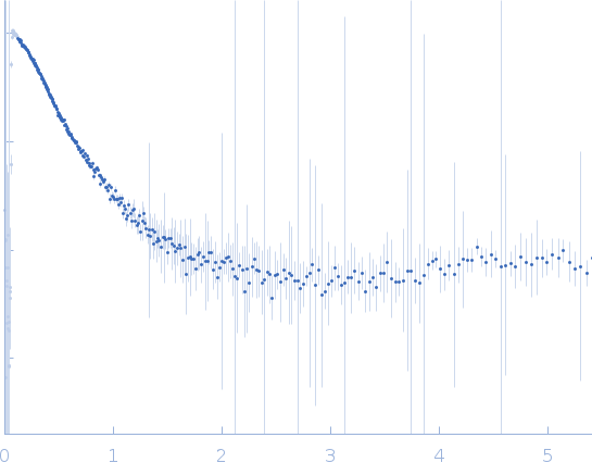
Histone H3.3 (H3.3)
Mol. type   Protein
Organism   Homo sapiens
Olig. state   Dimer
Mon. MW   4.2 kDa
 
UniProt   P84243 (1-40)
Sequence   FASTA
 
DNA methyltransferase 3 beta (215-853) (DNMT3B (215-853))
Mol. type   Protein
Organism   Homo sapiens
Olig. state   Dimer
Mon. MW   72.6 kDa
 
UniProt   Q9UBC3 (215-853)
Sequence   FASTA