| MWexperimental | 20 | kDa |
| MWexpected | 24 | kDa |
| VPorod | 28 | nm3 |
|
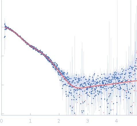
log I(s)
1.86×101
1.86×100
1.86×10⁻¹
1.86×10⁻²
|
 s, nm-1
|
|
|
|

|
|


|
|


|
|
Synchrotron SAXS
data from solutions of
S.aureus Protein Map Eap3 and Eap4 domains (Δ266-476)
in
20 mM HEPES, 140 mM NaCl, pH 7.4
were collected
on the
12.3.1 (SIBYLS) beam line
at the Advanced Light Source (ALS) storage ring
(Berkeley, CA, USA)
using a Pilatus3 X 2M detector
at a sample-detector distance of 2.1 m and
at a wavelength of λ = 0.1127 nm
(I(s) vs s, where s = 4πsinθ/λ, and 2θ is the scattering angle).
In-line size-exclusion chromatography (SEC) SAS was employed. The SEC parameters were as follows: A 60.00 μl sample
at 5 mg/ml was injected at a 0.65 ml/min flow rate
onto a Shodex KW-803 column
at 20°C.
660 successive
2 second frames were collected.
The data were normalized to the intensity of the transmitted beam and radially averaged; the scattering of the solvent-blank was subtracted.
|
|
|||||||||||||||||||||||||||