Ceylan B,
Adam J,
Toews S,
Kaiser F,
Dörr J,
Scheppa D,
Tants J,
Smart A,
Schoth J,
Philipp S,
Stirnal E,
Ferner J,
Richter C,
Sreeramulu S,
Caliskan N,
Schlundt A,
Weigand J,
Göbel M,
Wacker A,
Schwalbe H,
Angewandte Chemie International Edition
(2025)
DOI
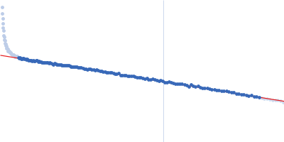
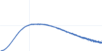
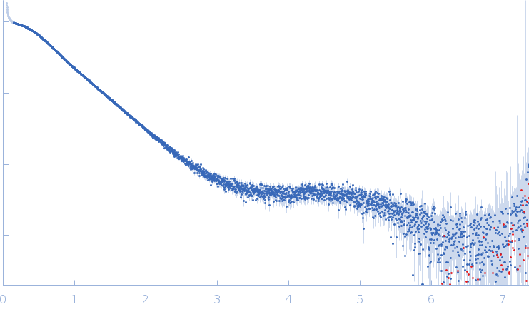
Synchrotron SAXS data from solutions of SARS-CoV2 RNA pseudoknot in 25 mM potassium phosphate, 50 mM KCl, pH 6.2 were collected on the EMBL P12 beam line at PETRA III (DESY; Hamburg, Germany) using a Pilatus 6M detector at a sample-detector distance of 3 m and at a wavelength of λ = 0.123982 nm (I(s) vs s, where s = 4πsinθ/λ, and 2θ is the scattering angle). One solute concentration of 4.00 mg/ml was measured at 20°C. 35 successive 0.095 second frames were collected. The data were normalized to the intensity of the transmitted beam and radially averaged; the scattering of the solvent-blank was subtracted.
Note: Possible repulsive interparticle interference present in the sample.
 s, nm-1