A glimpse into the hidden world of the flexible C-terminal protein binding domains of human RAD52

Struble L, Lovelace J, Borgstahl G, Journal of Structural Biology 216(3):108115 (2024) DOI

SASDVZ5 – Human DNA repair protein RAD52 homolog (amino acids 1-212)

DNA repair protein RAD52 homolog
MWexperimental 229 kDa
MWexpected 234 kDa
VPorod 375 nm3
log I(s) 8.03×100 8.03×10⁻¹ 8.03×10⁻² 8.03×10⁻³
DNA repair protein RAD52 homolog small angle scattering data  s, nm-1
ln I(s)
DNA repair protein RAD52 homolog Guinier plot ln 8.04×100 Rg: 4.1 nm 0 (4.1 nm)-2 s2
(sRg)2I(s)/I(0)
DNA repair protein RAD52 homolog Kratky plot 1.104 0 3 sRg
p(r)
DNA repair protein RAD52 homolog pair distance distribution function Rg: 4.0 nm 0 Dmax: 11.8 nm

Data validation


Fits and models


log I(s)
 s, nm-1
DNA repair protein RAD52 homolog DAMMIF model

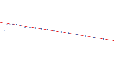
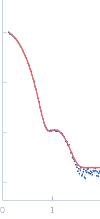
SAXS data from solutions of the human DNA repair protein RAD52 homolog (amino acids 1-212) in 20 mM Bis-Tris, 10% glycerol, 400 mM NaCl, 100 mM KCl, 1 mM EDTA, pH 6 were collected using a Rigaku/ BioSAXS1000 instrument at the Eppley Structural Biology Facility (University of Nebraska Medical Center, Omaha, NE, USA) equipped with a Dectris/Pilatus 100K-S detector at a sample-detector distance of 0.5 m and at a wavelength of λ = 0.154 nm (I(s) vs s, where s = 4πsinθ/λ, and 2θ is the scattering angle). One solute concentration of 7.48 mg/ml was measured at 15°C. One 9000 second frame was collected. The data were normalized to the intensity of the transmitted beam and radially averaged; the scattering of the solvent-blank was subtracted.

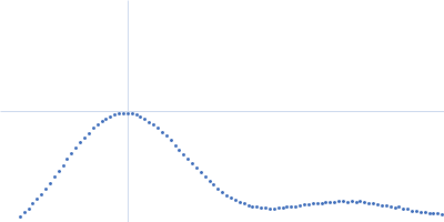
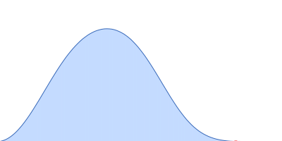
Using SEC-MALS the molecular weight is closest to that expected for a 10-mer. The data used to calculate the P(r) profile, and subsequent DAM modelling has been normalized to protein concentration in mg/mL.
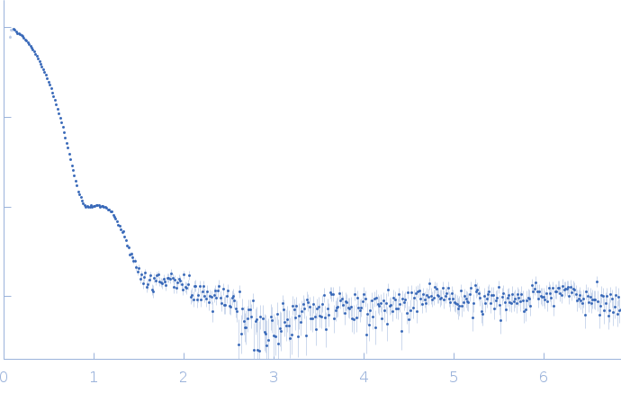
DNA repair protein RAD52 homolog (RAD52)
Mol. type   Protein
Organism   Homo sapiens
Olig. state   Decamer
Mon. MW   23.4 kDa
 
UniProt   P43351 (1-212)
Sequence   FASTA