|
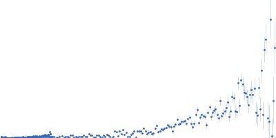
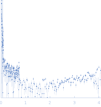
SANS data from solutions of β-chitin nanofibers from squid pens in 20 mM acetate, 47% v/v D₂O, pH 5 were collected on the D11 beam line at the Institut Laue-Langevin (ILL; Grenoble, France). Data recorded as I(s) vs s (where s = 4πsinθ/λ, and 2θ is the scattering angle), were collected at a sample concentration of 3 mg/mL at 25 degrees centigrade using three instrument configurations: Set-up 1: Sample-Detector distance, 1.7 m; Collimation distance, 5.5 m; Wavelength, 0.56 nm; Frames, 1; Time per frame, 1500 s. Set-up 2: Sample-Detector distance, 8.0 m; Collimation distance, 8.0 m; Wavelength, 0.56 nm; Frames, 1; Time per frame, 1500 s. Set-up 3: Sample-Detector distance, 38.95 m; Collimation distance, 40.0 m; Wavelength, 0.56 nm; Frames, 1; Time per frame, 2100 s.
Guinier analysis and subsequent Rg, I(0) and MW parameters are not applicable for this entry as the Guinier-region occurs at a much lower s-range than the data includes. The chitin fibers are micrometers long and are measured at the best-estimated neutron contrast match point. The match point is not perfect because the fibers form clusters with heterogeneous scattering length densities.
|
|
 s, nm-1