IgG4 and IgG1 undergo common acid‐induced compaction into an alternatively folded state

Imamura H, Honda S, FEBS Letters (2025) DOI

SASDW64 – Immunoglobulin G4 (pH 7)

humanized immunoglobulin G4 monoclonal antibody (lebrikizumab)
MWI(0) 146 kDa
MWexpected 148 kDa
VPorod 211 nm3
log I(s) 1.06×10⁻¹ 1.06×10⁻² 1.06×10⁻³ 1.06×10⁻⁴
humanized immunoglobulin G4 monoclonal antibody (lebrikizumab) small angle scattering data  s, nm-1
ln I(s)
humanized immunoglobulin G4 monoclonal antibody (lebrikizumab) Guinier plot ln 1.07×10⁻¹ Rg: 4.8 nm 0 (4.8 nm)-2 s2
(sRg)2I(s)/I(0)
humanized immunoglobulin G4 monoclonal antibody (lebrikizumab) Kratky plot 1.104 0 3 sRg
p(r)
humanized immunoglobulin G4 monoclonal antibody (lebrikizumab) pair distance distribution function Rg: 4.9 nm 0 Dmax: 15.6 nm

Data validation


There are no models related to this curve.

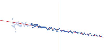
Synchrotron SAXS data from solutions of Immunoglobulin G4 in 10 mM sodium phosphate, pH 7 were collected on the BL-10C beam line at the Photon Factory High Energy Accelerator Research Organization (KEK; Tsukuba, Japan) using a Pilatus3 2M detector at a sample-detector distance of 1 m and at a wavelength of λ = 0.15 nm (I(s) vs s, where s = 4πsinθ/λ, and 2θ is the scattering angle). One solute concentration of 1.20 mg/ml was measured at 25°C. Five successive 2 second frames were collected. The data were normalized to the intensity of the transmitted beam and radially averaged; the scattering of the solvent-blank was subtracted.

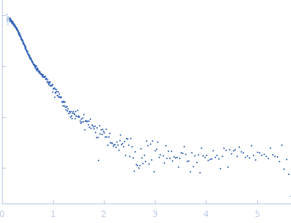
Minimum q reliable is ~0.15 nm-1, approximated by a standard sample (lysozyme protein) scattering. DrugBank ID: DB11914: https://go.drugbank.com/drugs/DB11914
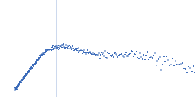
humanized immunoglobulin G4 monoclonal antibody (lebrikizumab) (IgG4)
Mol. type   Protein
Organism   Mouse/human
Olig. state   Monomer
Mon. MW   148 kDa
Sequence   FASTA