Tangled up in fibers: How a lytic polysaccharide monooxygenase binds its chitin substrate

Henrik Vinther Sørensen.

SASDW82 – Perdeuterated N-acetylglucosamine binding Protein A (D-GbpA) in 47 % D₂O, 2.4 mg/mL

GlcNAc-binding protein A (perdeuterated)
MWexperimental 44 kDa
MWexpected 51 kDa
log I(s) 1.22×10⁻¹ 1.22×10⁻² 1.22×10⁻³ 1.22×10⁻⁴
GlcNAc-binding protein A (perdeuterated) small angle scattering data  s, nm-1
ln I(s)
GlcNAc-binding protein A (perdeuterated) Guinier plot ln 1.23×10⁻¹ Rg: 3.7 nm 0 (3.7 nm)-2 s2
(sRg)2I(s)/I(0)
GlcNAc-binding protein A (perdeuterated) Kratky plot 1.104 0 3 sRg
p(r)
GlcNAc-binding protein A (perdeuterated) pair distance distribution function Rg: 3.7 nm 0 Dmax: 12.7 nm

Data validation


There are no models related to this curve.

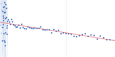
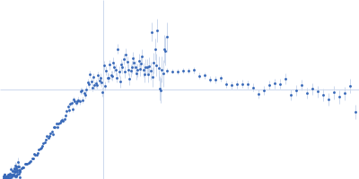
SANS data from solutions of perdeuterated N-acetylglucosamine binding Protein A (D-GbpA) in 100 mM NaCl, 20 mM Tris, 47% v/v D₂O, pH 8 were collected on the D11 beam line at the Institut Laue-Langevin (ILL; Grenoble, France). Data recorded as I(s) vs s (where s = 4πsinθ/λ, and 2θ is the scattering angle), were collected at a sample concentration of 2.4 mg/mL at 25 degrees centigrade using three instrument configurations: Set-up 1: Sample-Detector distance, 1.7 m; Collimation distance, 5.5 m; Wavelength, 0.56 nm; Frames, 1; Time per frame, 1500 s. Set-up 2: Sample-Detector distance, 8.0 m; Collimation distance, 8.0 m; Wavelength, 0.56 nm; Frames, 1; Time per frame, 1500 s. Set-up 3: Sample-Detector distance, 38.95 m; Collimation distance, 40.0 m; Wavelength, 0.56 nm; Frames, 1; Time per frame, 2100 s.
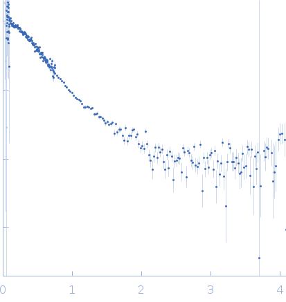
GlcNAc-binding protein A (perdeuterated) (D-GbpA)
Mol. type   Protein
Organism   Vibrio cholerae serotype O1 (strain ATCC 39315 / El Tor Inaba N16961)
Olig. state   Monomer
Mon. MW   51.3 kDa
 
UniProt   Q9KLD5 (24-485)
Sequence   FASTA