Sørensen HV,
Montserrat-Canals M,
Coder A,
Prévost S,
Krueger S,
Vaaje-Kolstad G,
Bjerregaard-Andersen K,
Lund R,
Krengel U,
ACS Appl Mater Interfaces
(2026)
Europe PMC
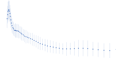
SASDWB2 – 3 mg/mL β-chitin nanofibers from squid pens with 2.5 mg/mL perdeuterated N-acetylglucosamine binding Protein A (D-GbpA) in 47 % D₂O - USANS
Dmax unknown – experimental data range validation not possible.
There are no models related to this curve.
Ultra small angle neutron scattering (USANS) data from of β-chitin nanofibers from squid pens mixed with perdeuterated N-acetylglucosamine binding Protein A (D-GbpA) in 20 mM acetate, 47% v/v D₂O, pH 5 were collected on the BT5 USANS instrument at the National Institute of Standards and Technology (NIST; Gaithersburg, MD, United States). Data recorded as I(s) vs s (where s = 4πsinθ/λ, and 2θ is the scattering angle), were collected at a sample concentration of 3 mg/mL chitin and 2.5 mg/mL D-GbpA at 25 degrees centigrade using the following instrument configuration: Sample-Detector distance, 30 m; Wavelength, 0.24 nm; Frames, 1; Time per frame, 11 hours; s-range, 0.0005 nm-1 to 0.01 nm-1.
Guinier analysis and subsequent Rg, I(0) and MW parameters are not applicable for this entry as the Guinier-region occurs at a much lower s-range than the data includes. The chitin fibers are micrometers long.
 s, nm-1