|
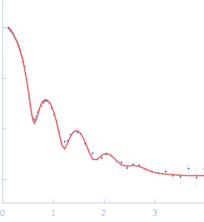
SANS data from solutions of apoferritin in 100 mM Tris-HCl, 100 mM NaCl in 100% v/v D₂O, pH 7.5 were collected on the SANS-U beam line at JRR-3 (Japan Atomic Energy Agency; Ibaraki, Japan) using a ORDELA, model 2660N detector at a sample-detector distance of 4 and 1.03 m at a wavelength of λ = 0.6 nm (I(s) vs s, where s = 4πsinθ/λ, and 2θ is the scattering angle). Stop-flow mode in-line size-exclusion chromatography (SEC) SAS was employed. The SEC parameters were as follows: A 500.00 μl sample at 12 mg/ml was injected onto a Cytiva Superdex 200 Increase 10/300 column at 25°C. 24 successive 600 second frames were collected. The data were normalized to the intensity of the transmitted beam and radially averaged; the scattering of the solvent-blank was subtracted.
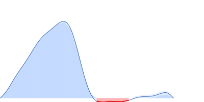
SEC elution paused during SANS measurements.
|
|
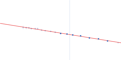
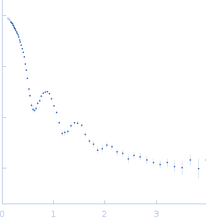 s, nm-1
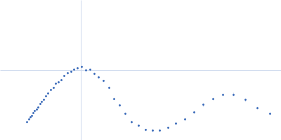