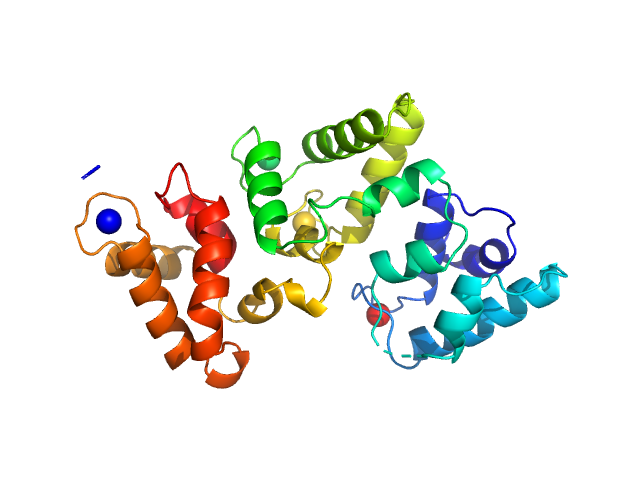
The X-ray structure of human calbindin-D28K: an improved model.
Noble JW,
Almalki R,
Roe SM,
Wagner A,
Duman R,
Atack JR
Acta Crystallogr D Struct Biol
74(Pt 10):1008-1014
(2018 Oct 1)
|
|
|
|
|
| Sample: |
Calbindin monomer, 30 kDa Homo sapiens protein
|
| Buffer: |
20 mM Tris, 150 mM NaCl, 3 mM CaCl2, pH: 7.8 |
| Experiment: |
SAXS
data collected at B21, Diamond Light Source on 2018 Feb 28
|
|
| RgGuinier |
2.1 |
nm |
| Dmax |
7.3 |
nm |
| VolumePorod |
47 |
nm3 |
|
|
|
|
|
|
|
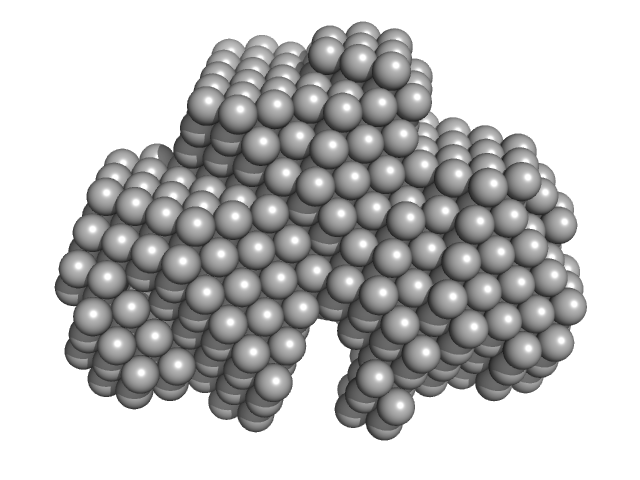
| Sample: |
Calbindin monomer, 30 kDa Homo sapiens protein
|
| Buffer: |
20 mM Tris, 150mM NaCl, pH: 7.8 |
| Experiment: |
SAXS
data collected at B21, Diamond Light Source on 2018 Feb 28
|
|
| RgGuinier |
2.1 |
nm |
| Dmax |
7.0 |
nm |
| VolumePorod |
46 |
nm3 |
|
|

