Characterization of the nucleotide-binding domain NsrF from the BceAB-type ABC-transporter NsrFP from the human pathogen Streptococcus agalactiae

Furtmann F, Porta N, Hoang D, Reiners J, Schumacher J, Gottstein J, Gohlke H, Smits S, Scientific Reports 10(1) (2020) DOI

SASDJR3 – BceAB-type ABC-Transporter nucleotide-binding domain, SaNsrF

ABC transporter, ATP-binding protein (Nucleotide-Binding Domain SaNsrF)
MWI(0) 32 kDa
MWexpected 31 kDa
VPorod 64 nm3
log I(s) 2.25×10⁻² 2.25×10⁻³ 2.25×10⁻⁴ 2.25×10⁻⁵
ABC transporter, ATP-binding protein (Nucleotide-Binding Domain SaNsrF) small angle scattering data  s, nm-1
ln I(s)
ABC transporter, ATP-binding protein (Nucleotide-Binding Domain SaNsrF) Guinier plot ln 2.25×10⁻² Rg: 2.4 nm 0 (2.4 nm)-2 s2
(sRg)2I(s)/I(0)
ABC transporter, ATP-binding protein (Nucleotide-Binding Domain SaNsrF) Kratky plot 1.104 0 3 sRg
p(r)
ABC transporter, ATP-binding protein (Nucleotide-Binding Domain SaNsrF) pair distance distribution function Rg: 2.5 nm 0 Dmax: 7.9 nm

Data validation


Fits and models


log I(s)
 s, nm-1
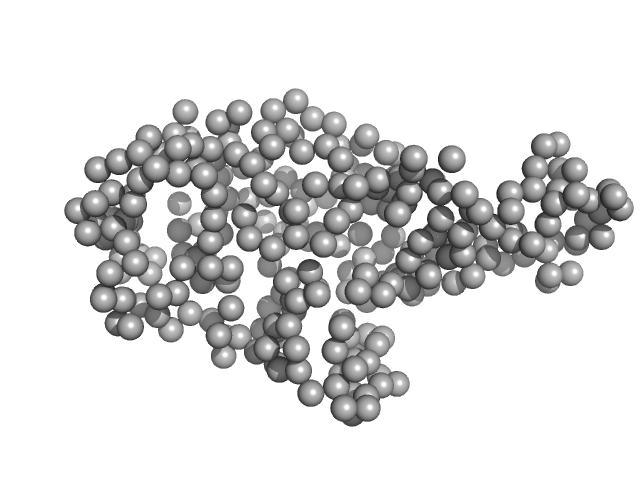
ABC transporter, ATP-binding protein (Nucleotide-Binding Domain SaNsrF) OTHER model

log I(s)
 s, nm-1
ABC transporter, ATP-binding protein (Nucleotide-Binding Domain SaNsrF) GASBOR model

SAXS data from solutions of SaNsrF in 100 mM HEPES, 150 mM NaCl, 10% glycerol, pH 8 were collected using a Xenocs Xeuss 2.0 Q-Zoom instrument equipped with a Pilatus3 R 300 K detector and a GENIX 3D CU Ultra Low Divergence X-ray beam delivery system (Center for Structural Studies, Heinrich-Heine-University, Düsseldorf, Germany). A sample-detector distance of 0.55 m was used and a X-ray wavelength of λ = 0.154 nm (I(s) vs s, where s = 4πsinθ/λ, and 2θ is the scattering angle). Solute concentrations ranging between 0.5 and 4.2 mg/ml were measured at 15°C. 12 successive 600 second frames were collected. The data were normalized to the intensity of the transmitted beam and radially averaged; the scattering of the solvent-blank was subtracted. The low angle data collected at lower concentration were merged with the highest concentration high angle data to yield the final composite scattering curve.

Samples were injected in the Low Noise Flow Cell (Xenocs) via autosampler. For each sample, twelve frames with an exposure time of ten minutes were collected. By comparing these frames, we excluded the possibility of aggregation and radiation damage during the measurement. Data were scaled to absolute intensity against water.

ABC transporter, ATP-binding protein (Nucleotide-Binding Domain SaNsrF) (SaNsrF)
Mol. type   Protein
Organism   Streptococcus agalactiae
Olig. state   Monomer
Mon. MW   30.9 kDa
 
UniProt   Q8DZX1 (1-250)
Sequence   FASTA