Solution structure, glycan specificity and of phenol oxidase inhibitory activity of Anopheles C-type lectins CTL4 and CTLMA2.

Bishnoi R, Sousa GL, Contet A, Day CJ, Hou CD, Profitt LA, Singla D, Jennings MP, Valentine AM, Povelones M, Baxter RHG, Sci Rep 9(1):15191 (2019) Europe PMC

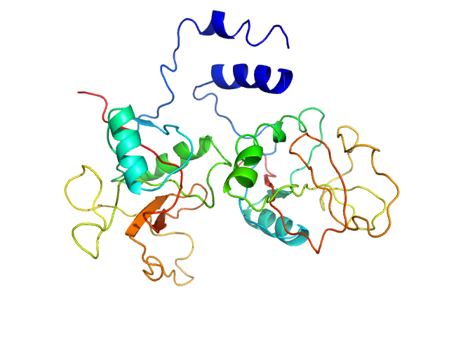
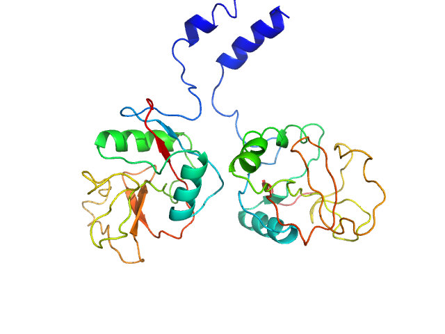
SASDFL4 – C-type lectins CTL4/CTLMA2, 3.1 mg/ml

AGAP005335-PA
AGAP005334-PA
MWexperimental 39 kDa
MWexpected 36 kDa
VPorod 58 nm3
log I(s) 6.50×10⁻¹ 6.50×10⁻² 6.50×10⁻³ 6.50×10⁻⁴
AGAP005335-PA AGAP005334-PA small angle scattering data  s, nm-1
ln I(s)
AGAP005335-PA AGAP005334-PA Guinier plot ln 6.50×10⁻¹ Rg: 2.5 nm 0 (2.5 nm)-2 s2
(sRg)2I(s)/I(0)
AGAP005335-PA AGAP005334-PA Kratky plot 1.104 0 3 sRg
p(r)
AGAP005335-PA AGAP005334-PA pair distance distribution function Rg: 2.5 nm 0 Dmax: 8 nm

Data validation


Fits and models


log I(s)
 s, nm-1
AGAP005335-PA AGAP005334-PA MULTIFOXS model

SAXS data from solutions of C-type lectins CTL4/CTLMA2 in 500 mM NaCl, 20 mM CHES, 0.5 mM CaCl2, 1% glycerol, pH 9 were collected on a Rigaku BioSAXS-2000 instrument at Thomas Jefferson University (Philadelphia, PA, USA) using a Pilatus 100K detector at a sample-detector distance of 0.481 m and at a wavelength of λ = 0.154187 nm (l(s) vs s, where s = 4πsinθ/λ, and 2θ is the scattering angle). One solute concentration of 3.10 mg/ml was measured at 20°C. Six successive 300 second frames were collected. The data were normalized to the intensity of the transmitted beam and radially averaged; the scattering of the solvent-blank was subtracted.

AGAP005335-PA (CTL4)
Mol. type   Protein
Organism   Anopheles gambiae
Olig. state   Monomer
Mon. MW   18.1 kDa
 
UniProt   Q7Q7F7 (25-177)
Sequence   FASTA
 
AGAP005334-PA (CTLMA2)
Mol. type   Protein
Organism   Anopheles gambiae
Olig. state   Monomer
Mon. MW   17.9 kDa
 
UniProt   Q7Q7F8 (18-174)
Sequence   FASTA