Solution Structure and Conformational Dynamics of a Doublet Acyl Carrier Protein from Prodigiosin Biosynthesis.

Thongkawphueak T, Winter AJ, Williams C, Maple HJ, Soontaranon S, Kaewhan C, Campopiano DJ, Crump MP, Wattana-Amorn P, Biochemistry (2021) Europe PMC

SASDDH6 – Di-domain acyl carrier protein of the seryltransferase from prodigiosin biosynthesis

Di-domain acyl carrier protein of PigH from prodigiosin biosynthesis
MWexperimental 19 kDa
MWexpected 22 kDa
VPorod 39 nm3
log I(s) 2.27×10⁻⁵ 2.27×10⁻⁶ 2.27×10⁻⁷ 2.27×10⁻⁸
Di-domain acyl carrier protein of PigH from prodigiosin biosynthesis small angle scattering data  s, nm-1
ln I(s)
Di-domain acyl carrier protein of PigH from prodigiosin biosynthesis Guinier plot ln 2.28×10⁻⁵ Rg: 2.4 nm 0 (2.4 nm)-2 s2
(sRg)2I(s)/I(0)
Di-domain acyl carrier protein of PigH from prodigiosin biosynthesis Kratky plot 1.104 0 3 sRg
p(r)
Di-domain acyl carrier protein of PigH from prodigiosin biosynthesis pair distance distribution function Rg: 2.6 nm 0 Dmax: 8.4 nm

Data validation


Fits and models


log I(s)
 s, nm-1
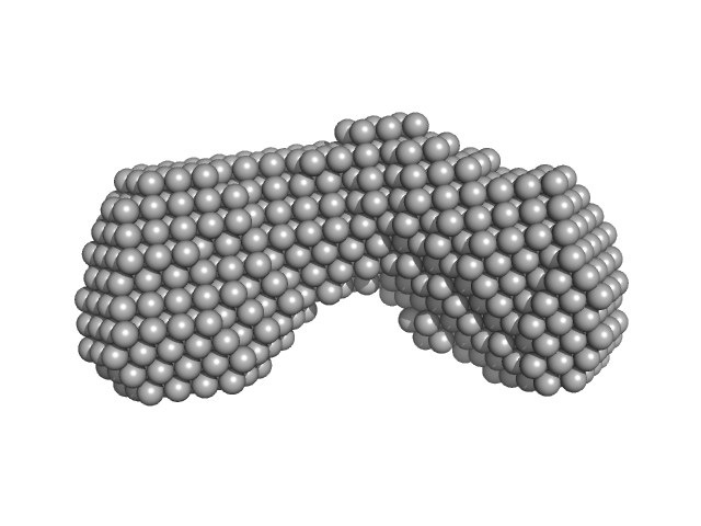
Di-domain acyl carrier protein of PigH from prodigiosin biosynthesis DAMMIF model
Di-domain acyl carrier protein of PigH from prodigiosin biosynthesis DAMFILT model

Synchrotron SAXS data from solutions of the di-domain acyl carrier protein of the seryltransferase from prodigiosin biosynthesis in 20 mM Tris, 5 mM DTT, pH 7, were collected on the BL1.3W beam line at the Synchrotron Light Research Institute (Nakhon Ratchasima, Thailand) using a CCD Rayonix SX165 detector at a sample-detector distance of 0.9 m and at a wavelength of λ = 0.131 nm (l(s) vs s, where s = 4πsinθ/λ, and 2θ is the scattering angle). Solute concentrations ranging between 1.7 and 6.8 mg/ml were measured at 25°C. One 600 second frame was collected. The data were normalized to the intensity of the transmitted beam and radially averaged; the scattering of the solvent-blank was subtracted. The low angle data collected at lower concentration were merged with the highest concentration high angle data to yield the final composite scattering curve.

Two DAMMIF-derived dummy atom models are displayed. Top: The best-fit individual DAMMIF model and corresponding fit to the SAXS data. Bottom: The average spatial representation of the protein obtained after the alignment of multiple individual DAMMIF models combined with volume and bead-occupancy corrections (damfilt).

Di-domain acyl carrier protein of PigH from prodigiosin biosynthesis (Di-domain ACP)
Mol. type   Protein
Organism   Serratia sp. ATCC 39006
Olig. state   Monomer
Mon. MW   22.2 kDa
 
UniProt   Q5W264 (4-191)
Sequence   FASTA