Leucine Rich Repeat And Sterile Alpha Motif Containing 1 (LRSAM1)

Si Hoon Park.

SASDMR3 – Wild type E3 ubiquitin-protein ligase LRSAM1 (561-632): SAM domain

E3 ubiquitin-protein ligase LRSAM1 - SAM domain
MWexperimental 10 kDa
MWexpected 8 kDa
VPorod 11 nm3
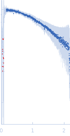
log I(s) 4.54×100 4.54×10⁻¹ 4.54×10⁻² 4.54×10⁻³
E3 ubiquitin-protein ligase LRSAM1 - SAM domain small angle scattering data  s, nm-1
ln I(s)
E3 ubiquitin-protein ligase LRSAM1 - SAM domain Guinier plot ln 4.55×100 Rg: 1.4 nm 0 (1.4 nm)-2 s2
(sRg)2I(s)/I(0)
E3 ubiquitin-protein ligase LRSAM1 - SAM domain Kratky plot 1.104 0 3 sRg
p(r)
E3 ubiquitin-protein ligase LRSAM1 - SAM domain pair distance distribution function Rg: 1.4 nm 0 Dmax: 4.3 nm

Data validation


Fits and models


log I(s)
 s, nm-1
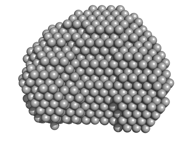
E3 ubiquitin-protein ligase LRSAM1 - SAM domain DAMFILT model
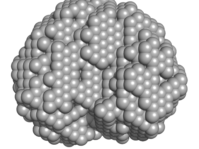
E3 ubiquitin-protein ligase LRSAM1 - SAM domain DAMMIN model

Synchrotron SAXS data from solutions of LRSAM1 (amino acids 561-632) in 50 mM Tris-HCl pH 8.0, 150 mM NaCl, 1 mM TCEP, pH 8 were collected on the 4C beam line at the Pohang Accelerator Laboratory (Pohang, South Korea) using a Rayonix SX165 detector at a sample-detector distance of 3 m and at a wavelength of λ = 0.0734 nm (I(s) vs s, where s = 4πsinθ/λ, and 2θ is the scattering angle). One solute concentration of 16.20 mg/ml was measured at 20°C. Six successive 10 second frames were collected. The data were normalized to the intensity of the transmitted beam and radially averaged; the scattering of the solvent-blank was subtracted.

The models displayed show both the average-weighted bead-occupancy and volume-corrected representation of the protein calculated from the spatial alignment of a cohort of individual models (damfilt; top) and an individual model reconstruction (bottom) with the associated fit to the data.

E3 ubiquitin-protein ligase LRSAM1 - SAM domain (LRSAM1)
Mol. type   Protein
Organism   Homo sapiens
Olig. state   Monomer
Mon. MW   8.2 kDa
 
UniProt   Q6UWE0 (561-632)
Sequence   FASTA