Development of DNA Aptamers for Visualization of Glial Brain Tumors and Detection of Circulating Tumor Cells

Kichkailo A, Narodov A, Komarova M, Zamay T, Zamay G, Kolovskaya O, Erakhtin E, Glazyrin Y, Veprintsev D, Moryachkov R, Zabluda V, Shchugoreva I, Artyushenko P, Mironov V, Morozov D, Khorzhevskii V, Gorbushin A, Koshmanova A, Nikolaeva E, Grinev I, Voronkovskii I, Grek D, Belugin K, Volzhentsev A, Badmaev O, Luzan N, Lukyanenko K, Peters G, Lapin I, Kirichenko A, Konarev P, Morozov E, Mironov G, Gargaun A, Muharemagic D, Zamay S, Kochkina E, Dymova M, Smolyarova T, Sokolov A, Modestov A, Tokarev N, Shepelevich N, Ozerskaya A, Chanchikova N, Krat A, Zukov R, Bakhtina V, Shnyakin P, Shesternya P, Svetlichnyi V, Petrova M, Artyukhov I, Tomilin F, Berezovski M, Molecular Therapy - Nucleic Acids (2023) DOI

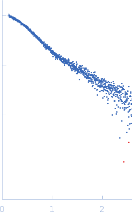
SASDP29 – Gli-55 - ssDNA aptamer specific to glioma brain tumor cells

Gli-55 - ssDNA aptamer specific to glioma brain tumor cells
MWexperimental 19 kDa
MWexpected 19 kDa
VPorod 34 nm3
log I(s) 1.05×10³ 1.05×10² 1.05×101 1.05×100
Gli-55 - ssDNA aptamer specific to glioma brain tumor cells small angle scattering data  s, nm-1
ln I(s)
Gli-55 - ssDNA aptamer specific to glioma brain tumor cells Guinier plot ln 1.06×10³ Rg: 2.8 nm 0 (2.8 nm)-2 s2
(sRg)2I(s)/I(0)
Gli-55 - ssDNA aptamer specific to glioma brain tumor cells Kratky plot 1.104 0 3 sRg
p(r)
Gli-55 - ssDNA aptamer specific to glioma brain tumor cells pair distance distribution function Rg: 2.9 nm 0 Dmax: 11.5 nm

Data validation


Fits and models


log I(s)
 s, nm-1
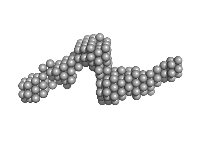
Gli-55 - ssDNA aptamer specific to glioma brain tumor cells DAMMIN model

log I(s)
 s, nm-1
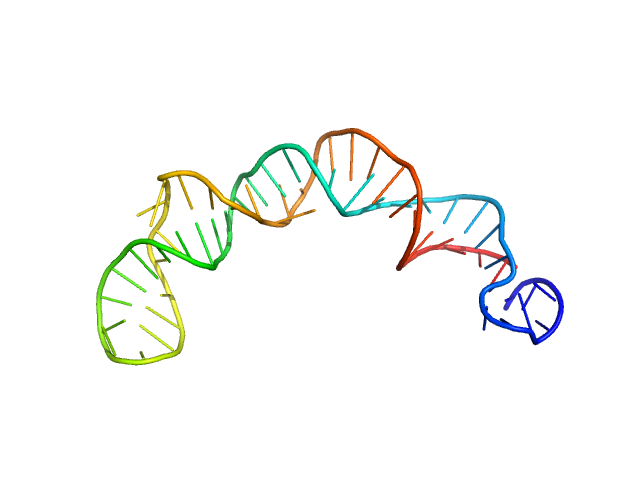
Gli-55 - ssDNA aptamer specific to glioma brain tumor cells GROMACS model

SAXS data for the Gli-55 aptamer were obtained at the P12 BioSAXS beamline (EMBL) at the Petra III storage ring of the synchrotron center DESY, Hamburg. The eluent of the employed chromatography column was passed through a 1.7 mm quartz capillary held under vacuum. The SAXS data were recorded on a Pilatus 6M area detector (Dectris) at a sample to detector distance of 3 meters and the wavelength λ = 0.124 nm (X-ray energy 10 keV) at room temperature 20.4 °C. Series of individual 1 s exposure X-ray data frames were measured from the continuously-flowing column eluate across one column volume. The two-dimensional SAXS intensities were reduced to I(s) vs s using the integrated analysis pipeline SASFLOW. The s-axis was calibrated with silver behenate and the resulting profiles were normalized for exposure time and sample transmission. To distinguish the oligomeric constituents in solution the joint technique of size-exclusion chromatography with SAXS (SEC-SAXS) was applied. To decompose the partially overlapping components in SEC-SAXS data the evolving factor analysis (EFA) was applied using the program EFAMIX.

Gli-55 - ssDNA aptamer specific to glioma brain tumor cells (Gli-55)
Mol. type   DNA
Organism   Artificially synthesized
Olig. state   Monomer
Mon. MW   18.6 kDa
Sequence   FASTA