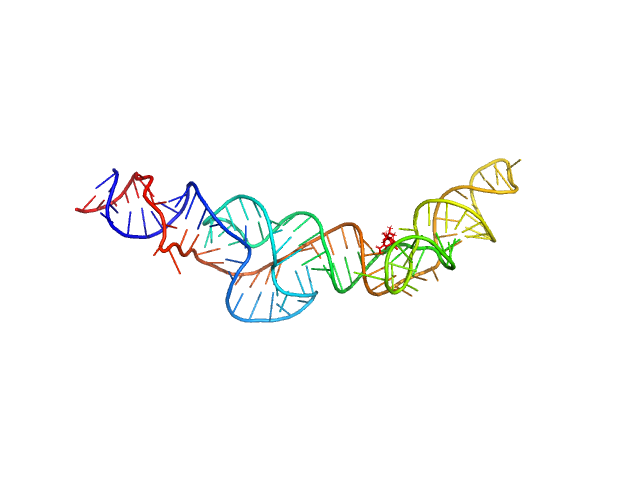
Structural investigation of an RNA device that regulates PD-1 expression in mammalian cells.

Stagno JR, Deme JC, Dwivedi V, Lee YT, Lee HK, Yu P, Chen SY, Fan L, Degenhardt MFS, Chari R, Young HA, Lea SM, Wang YX, Nucleic Acids Res 53(5) (2025) Europe PMC

SASDSQ8 – RNA Device 43 in ligand-bound state (holo-D43)

RNA Device 43 in ligand-bound state
MWexperimental 39 kDa
MWexpected 40 kDa
VPorod 56 nm3
log I(s) 2.47×101 2.47×100 2.47×10⁻¹ 2.47×10⁻²
RNA Device 43 in ligand-bound state small angle scattering data  s, nm-1
ln I(s)
RNA Device 43 in ligand-bound state Guinier plot ln 2.47×101 Rg: 3.3 nm 0 (3.3 nm)-2 s2
(sRg)2I(s)/I(0)
RNA Device 43 in ligand-bound state Kratky plot 1.104 0 3 sRg
p(r)
RNA Device 43 in ligand-bound state pair distance distribution function Rg: 3.5 nm 0 Dmax: 12.1 nm

Data validation


Fits and models


log I(s)
 s, nm-1
RNA Device 43 in ligand-bound state OTHER model

Synchrotron SAXS data from solutions of holo-D43 in 10 mM Bis-Tris, 100 mM KCl, and 1 mM MgCl2, pH 6.8 were collected on the 16-ID (LiX) beam line at the National Synchrotron Light Source II (NSLS-II; Upton, NY, USA) using a Pilatus3 S 1M detector at a sample-detector distance of 3.7 m and at a wavelength of λ = 0.08202 nm (I(s) vs s, where s = 4πsinθ/λ, and 2θ is the scattering angle). Solute concentrations ranging between 1.2 and 10 mg/ml were measured at 20°C. 20 successive 0.500 second frames were collected. The data were normalized to the intensity of the transmitted beam and radially averaged; the scattering of the solvent-blank was subtracted. The low angle data collected at lower concentrations were extrapolated to infinite dilution and merged with the higher concentration data to yield the final composite scattering curve.

The MW for the RNA was calculate using the volume of correlation method.

RNA Device 43 in ligand-bound state (holo-D43)
Mol. type   RNA
Organism   synthetic construct
Olig. state   Monomer
Mon. MW   40.2 kDa
Sequence   FASTA