A Highly Conserved Yet Flexible Linker Is Part of a Polymorphic Protein-Binding Domain in Myosin-Binding Protein C.

Michie KA, Kwan AH, Tung CS, Guss JM, Trewhella J, Structure 24(11):2000-2007 (2016) Europe PMC

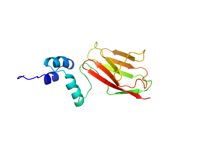
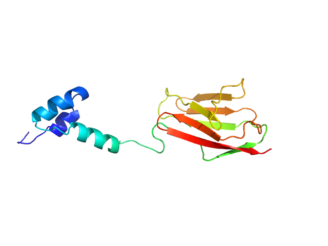
SASDDD9 – Cardiac myosin binding protein C: tri-helix bundle from the motif with C2 domain

cardiac myosin binding protein C: tri-helix bundle-C2
MWI(0) 18 kDa
MWexpected 15 kDa
VPorod 20 nm3
log I(s) 4.60×10⁻² 4.60×10⁻³ 4.60×10⁻⁴ 4.60×10⁻⁵
cardiac myosin binding protein C: tri-helix bundle-C2 small angle scattering data  s, nm-1
ln I(s)
cardiac myosin binding protein C: tri-helix bundle-C2 Guinier plot ln 4.60×10⁻² Rg: 1.9 nm 0 (1.9 nm)-2 s2
(sRg)2I(s)/I(0)
cardiac myosin binding protein C: tri-helix bundle-C2 Kratky plot 1.104 0 3 sRg
p(r)
cardiac myosin binding protein C: tri-helix bundle-C2 pair distance distribution function Rg: 2.0 nm 0 Dmax: 8 nm

Data validation


Fits and models


log I(s)
 s, nm-1
Cardiac myosin binding protein C: tri-helix bundle from the motif with C2 domain Rg histogram Rg, nm

log I(s)
 s, nm-1
cardiac myosin binding protein C: tri-helix bundle-C2 MULTIFOXS model

Synchrotron SAXS data from solutions of the cardiac myosin binding protein C tri-helixmotif bundle and C2 domain in 150 mM NaCl, 10 mM MES, 2 mM TCEP, 1 mM NaN3, pH 6.5 (at 4°C) were collected at the SAXS/WAXS beam line at the Australian Synchrotron (Melbourne, Australia) using a Pilatus 1M detector at a sample-detector distance of 2.7 m and at a wavelength of λ = 0.10332 nm (l(s) vs s, where s = 4πsinθ/λ, and 2θ is the scattering angle). Solute concentrations ranging between 1.0 and 4.9 mg/ml were measured in batch mode. Data were reduced to I(s) versus s using the software ScatterBrain (http://www.synchrotron.org.au/aussyncbeamlines/saxswaxs/software-saxswaxs). Intensities were placed on an absolute scale using the known scattering from H2O. Samples were prepared by gel fitration with a solvent scattering blank taken from buffer eluted from the gel filtration column was measured before each sample. Scattering profiles were obtained by subtraction of the solvent blank scattering from the protein+solvent scattering and extrapolated to zero concentration in primus qt.

The concentration series SAXS data and EOM models are provided in the full entry zip archive.

cardiac myosin binding protein C: tri-helix bundle-C2 (tri-helix bundle-C2)
Mol. type   Protein
Organism   human sequence obtained using E. coli expression
Olig. state   Monomer
Mon. MW   15.5 kDa
 
UniProt   Q14896 (319-451)
Sequence   FASTA