Structural Contour Map of the Iota Carbonic Anhydrase from the Diatom Thalassiosira pseudonana Using a Multiprong Approach.

Jensen EL, Receveur-Brechot V, Hachemane M, Wils L, Barbier P, Parsiegla G, Gontero B, Launay H, Int J Mol Sci 22(16) (2021) Europe PMC

SASDMM2 – Thalassiosira pseudonana iota Carbonic anhydrase (ι-CA)

iota carbonic anhydrase
MWexperimental 292 kDa
MWexpected 260 kDa
VPorod 500 nm3
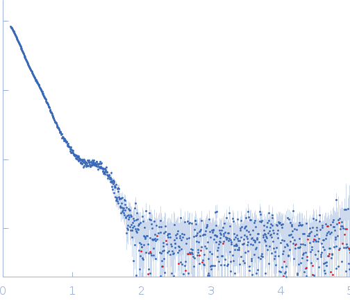
log I(s) 1.01×10⁻¹ 1.01×10⁻² 1.01×10⁻³ 1.01×10⁻⁴
iota carbonic anhydrase small angle scattering data  s, nm-1
ln I(s)
iota carbonic anhydrase Guinier plot ln 1.01×10⁻¹ Rg: 6.7 nm 0 (6.7 nm)-2 s2
(sRg)2I(s)/I(0)
iota carbonic anhydrase Kratky plot 1.104 0 3 sRg
p(r)
iota carbonic anhydrase pair distance distribution function Rg: 6.8 nm 0 Dmax: 25 nm

Data validation


Fits and models


log I(s)
 s, nm-1
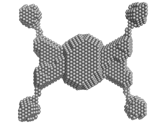
iota carbonic anhydrase DAMMIF model

Synchrotron SAXS data from solutions of Thalassiosira pseudonana ι-CA in 20 mM Tris, 150 mM NaCl, pH 8 were collected on the SWING beam line at SOLEIL (Saint-Aubin, France) using a Eiger 4M detector at a sample-detector distance of 2 m and at a wavelength of λ = 0.1 nm (I(s) vs s, where s = 4πsinθ/λ, and 2θ is the scattering angle). In-line size-exclusion chromatography (SEC) SAS was employed. The SEC parameters were as follows: A 50.00 μl sample at 4.8 mg/ml was injected at a 0.30 ml/min flow rate onto a column at 15°C. 500 successive 0.990 second frames were collected. The data were normalized to the intensity of the transmitted beam and radially averaged; the scattering of the solvent-blank was subtracted from the main SEC-elution peak data frames, corresponding to the protein tetramer.

SEC column = UNKNOWN

iota carbonic anhydrase (ι-CA)
Mol. type   Protein
Organism   Thalassiosira pseudonana
Olig. state   Tetramer
Mon. MW   65.1 kDa
Sequence   FASTA