Ferritin-based fusion protein shows octameric deadlock state of self-assembly

Sudarev V, Gette M, Bazhenov S, Tilinova O, Zinovev E, Manukhov I, Kuklin A, Ryzhykau Y, Vlasov A, Biochemical and Biophysical Research Communications 690:149276 (2024) DOI

SASDTW4 – Ferritin from Helicobacter pylori with an N-terminal His6-tag

Bacterial non-heme ferritin (N19Q, I59V)
MWexperimental 480 kDa
MWexpected 480 kDa
VPorod 674 nm3
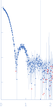
log I(s) 4.37×100 4.37×10⁻¹ 4.37×10⁻² 4.37×10⁻³
Bacterial non-heme ferritin (N19Q, I59V) small angle scattering data  s, nm-1
ln I(s)
Bacterial non-heme ferritin (N19Q, I59V) Guinier plot ln 4.38×100 Rg: 5.6 nm 0 (5.6 nm)-2 s2
(sRg)2I(s)/I(0)
Bacterial non-heme ferritin (N19Q, I59V) Kratky plot 1.104 0 3 sRg
p(r)
Bacterial non-heme ferritin (N19Q, I59V) pair distance distribution function Rg: 5.4 nm 0 Dmax: 14.5 nm

Data validation


Fits and models


log I(s)
 s, nm-1
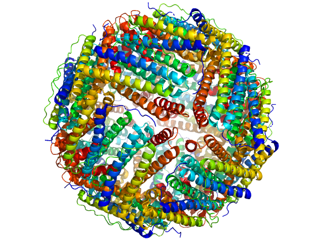
Bacterial non-heme ferritin (N19Q, I59V) PDB (PROTEIN DATA BANK) model

SAXS data from solutions of ferritin in 20 mM Tris, pH 8 were collected using a Rigaku MicroMax 007-HF instrument at the Moscow Institute of Physics and Technology (MIPT; Dolgoprudny, Russian Federation) equipped with a multiwire gas-filled ASM DTR Triton 200 detector at a sample-detector distance of 2 m and at a wavelength of λ = 0.15406 nm (I(s) vs s, where s = 4πsinθ/λ, and 2θ is the scattering angle). One solute concentration of 20.00 mg/ml was measured at 20°C. One 7980 second frame was collected. The data were normalized to the intensity of the transmitted beam and radially averaged; the scattering of the solvent-blank was subtracted.

Bacterial non-heme ferritin (N19Q, I59V) (FerHis)
Mol. type   Protein
Organism   Helicobacter pylori (strain J99 / ATCC 700824)
Olig. state   24-mer
Mon. MW   20.0 kDa
 
UniProt   Q9ZLI1 (5-167)
Sequence   FASTA
 
PDB ID   3BVE