Insight into the structure and interactions of the M. tuberculosis Mce-associated membrane proteins Mam1A-1D

Hynönen M, Perumal P, Hynönen N, Doutch J, Ma K, Venkatesan R, (2025) DOI

SASDXG2 – Probable conserved Mce associated membrane protein (Mam1Adelta106) from M.tuberculosis (SANS)

Probable conserved Mce associated membrane protein
MWexperimental 33 kDa
MWexpected 59 kDa
VPorod 33 nm3
log I(s) 1.24×10⁻¹ 1.24×10⁻² 1.24×10⁻³ 1.24×10⁻⁴
Probable conserved Mce associated membrane protein small angle scattering data  s, nm-1
ln I(s)
Probable conserved Mce associated membrane protein Guinier plot ln 1.24×10⁻¹ Rg: 2.6 nm 0 (2.6 nm)-2 s2
(sRg)2I(s)/I(0)
Probable conserved Mce associated membrane protein Kratky plot 1.104 0 3 sRg
p(r)
Probable conserved Mce associated membrane protein pair distance distribution function Rg: 2.6 nm 0 Dmax: 8.5 nm

Data validation


Fits and models


log I(s)
 s, nm-1
Probable conserved Mce associated membrane protein DAMFILT model
Probable conserved Mce associated membrane protein ALPHAFOLD model

log I(s)
 s, nm-1
Probable conserved Mce associated membrane protein DAMFILT model
Probable conserved Mce associated membrane protein ALPHAFOLD model

SANS 
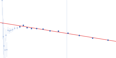
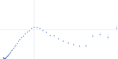
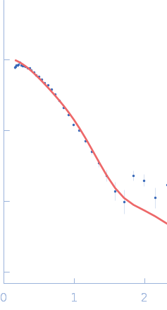
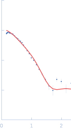
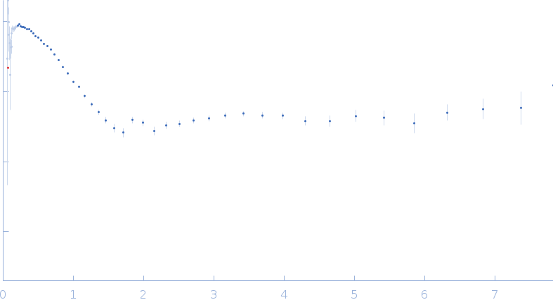
data from solutions of Probable conserved Mce associated membrane protein (Mam1Adelta106) from M.tuberculosis (SANS) in 50 mM Tris, 500 mM NaCl, D-C12E9, pH 8.5 were collected (I(s) vs s, where s = 4πsinθ/λ, and 2θ is the scattering angle). at 6°C. The data were normalized to the intensity of the transmitted beam and radially averaged; the scattering of the solvent-blank was subtracted.

SANS data were collected on the Zoom time-of-flight SANS instrument (ISIS Neutron and Muon Source, Harwell, UK) using wavelength range 1.75-16.5Å. The source to sample and sample to detector distance was 4 m and the beam size was 12 mm. Mam1Adelta106 was solubilized with C12E9 in the lysis buffer where the deuteration of the C12E9 was 0.2 % v/v. Excess C12E9 was washed away throughout the purification, and replaced with D-C12E9 in the STFC SANS facilities using SEC Superdex200 column. From the highest point of the peak, 3 fractions were selected. Each of these fractions was loaded to Starna Scientific quartz round cuvette (match code 6,path length 1, type 32/Q/1), and SANS data were collected from each fraction with an exposure time of 1 h in 5 cycles (5 h total exposure time for each fraction). The neutron momentum transfer (q) standard deviation is provided as an additional file.

Probable conserved Mce associated membrane protein (Mam1A delta 106)
Mol. type   Protein
Organism   Mycobacterium tuberculosis (strain ATCC 25618 / H37Rv)
Olig. state   Tetramer
Mon. MW   14.8 kDa
 
UniProt   O07419 (107-213)
Sequence   FASTA